• 文献检索
  • 文档翻译
  • 深度研究
  • 学术资讯
  • Suppr Zotero 插件Zotero 插件
  • 邀请有礼
  • 套餐&价格
  • 历史记录
应用&插件
Suppr Zotero 插件Zotero 插件浏览器插件Mac 客户端Windows 客户端微信小程序
定价
高级版会员购买积分包购买API积分包
服务
文献检索文档翻译深度研究API 文档MCP 服务
关于我们
关于 Suppr公司介绍联系我们用户协议隐私条款
关注我们

Suppr 超能文献

核心技术专利:CN118964589B侵权必究
粤ICP备2023148730 号-1Suppr @ 2026

文献检索

告别复杂PubMed语法,用中文像聊天一样搜索,搜遍4000万医学文献。AI智能推荐,让科研检索更轻松。

立即免费搜索

文件翻译

保留排版,准确专业,支持PDF/Word/PPT等文件格式,支持 12+语言互译。

免费翻译文档

深度研究

AI帮你快速写综述,25分钟生成高质量综述,智能提取关键信息,辅助科研写作。

立即免费体验

经动脉化疗栓塞后肝细胞癌残余肿瘤的超声造影:有助于肿瘤反应评估吗?

Contrast-Enhanced Ultrasound in Residual Tumor of Hepatocellular Carcinoma following Transarterial Chemoembolization: Is It Helpful for Tumor Response?

机构信息

Department of Ultrasound, The First Affiliated Hospital, Medical College, Zhejiang University, Hangzhou, China.

Key Laboratory of Precision Diagnosis and Treatment for Hepatobiliary and Pancreatic Tumor of Zhejiang Province, Hangzhou 310003, China.

出版信息

Biomed Res Int. 2018 Aug 2;2018:8632069. doi: 10.1155/2018/8632069. eCollection 2018.

DOI:10.1155/2018/8632069
PMID:30175147
原文链接:https://pmc.ncbi.nlm.nih.gov/articles/PMC6098931/
Abstract

AIM

To investigate the enhancement pattern of residual tumor on contrast-enhanced ultrasonography (CEUS) in patients with hepatocellular carcinoma (HCC) treated with transarterial chemoembolization (TACE).

METHODS

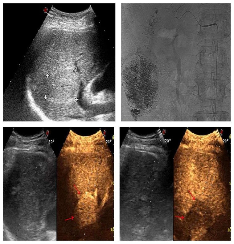
Our study initially included 76 patients with HCC, 73 of which were finally allocated into two groups: group 1 (43 patients, post-TACE group) and group 2 (30 patients, untreated HCC group). All patients were performed with CEUS using SonoVue, and qualitative and quantitative enhancement characteristics (rise time, peak time, and washout time) were evaluated for the residual tumors. T test or 2 test was used to estimate for differences between two groups.

RESULTS

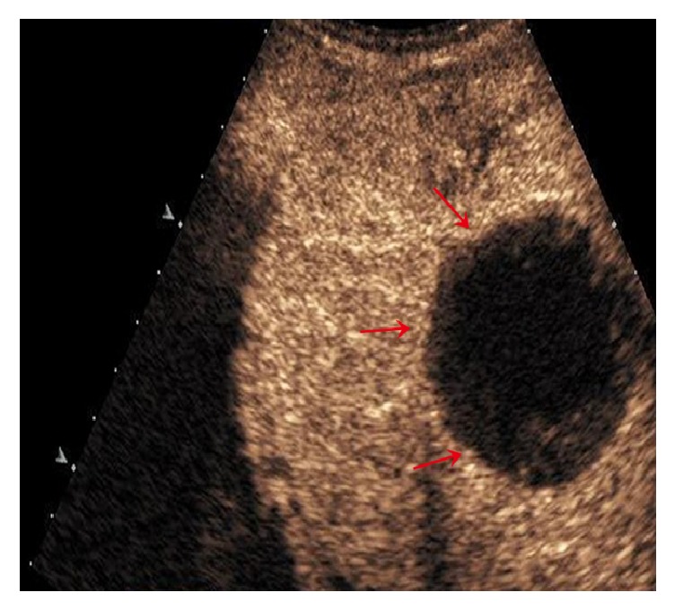
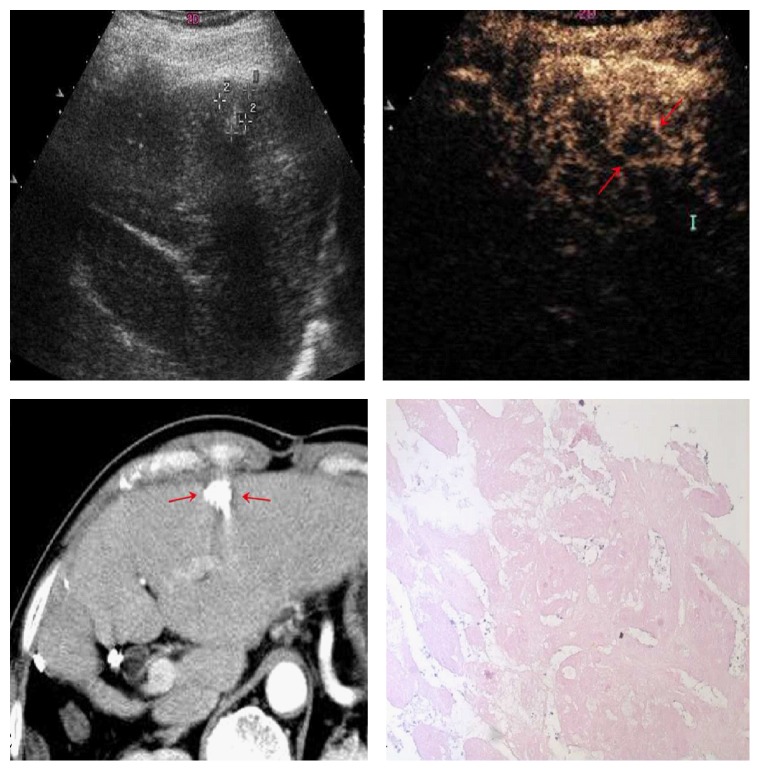
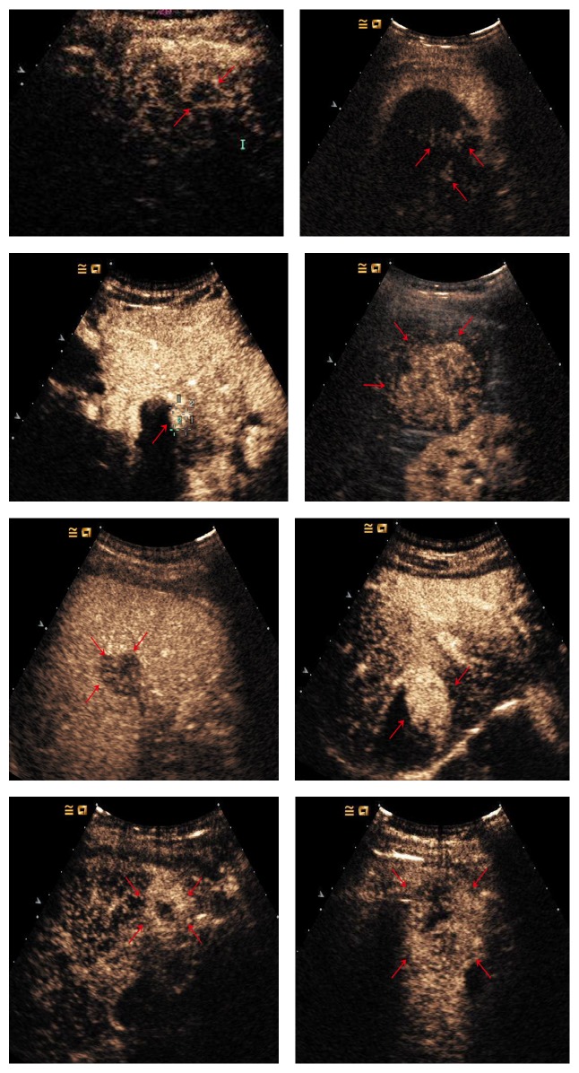
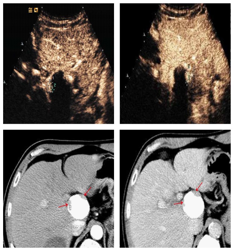
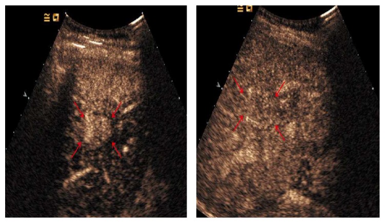
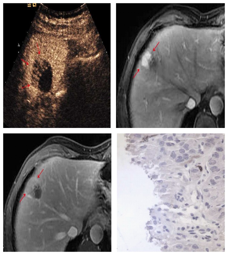
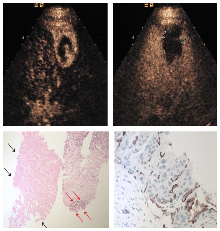
In group 1, the mean rise time, peak time, and washout times in group 1 were 16.1±2.7 sec, 31.3±3.1 sec, and 191.0±31.3 sec, respectively. In group 2, these were 15.1±3.5 sec, 30.9±3.2 sec, and 142.6±16.1 sec, respectively. The differences in rise time and peak time were not statistically significant (=0.09 and 0.30, respectively), but the washout time was significantly prolonged in group 1 (<0.01). The enhanced pattern in arterial phase was inhomogeneous (n=11), regular homogeneous (n=11), partial (n=12), peripheral (n=7), and peripheral rim-like (n=2) in group 1. The average of the longest tumor size of the whole lesion in the 5 types was 4.7±1.3cm, 2.9±1.0cm, 3.1±1.7cm, 2.5±0.6cm, and 2.1 cm.

CONCLUSION

It suggested that the washout time of post-TACE residual lesions was prolonged compared with untreated HCC nodules on CEUS imaging. Combined with the triple-phase enhancement pattern seen on CEUS, the washout time may provide additional information to guide further treatment for residual tumors.

摘要

目的

探讨经导管肝动脉化疗栓塞(TACE)治疗后肝细胞癌(HCC)患者残留肿瘤在超声造影(CEUS)中的增强模式。

方法

本研究最初纳入 76 例 HCC 患者,其中 73 例最终分为两组:组 1(43 例,TACE 后组)和组 2(30 例,未治疗 HCC 组)。所有患者均采用 SonoVue 进行 CEUS,评估残留肿瘤的定性和定量增强特征(上升时间、峰值时间和洗脱时间)。采用 t 检验或卡方检验估计两组间差异。

结果

在组 1 中,组 1 的平均上升时间、峰值时间和洗脱时间分别为 16.1±2.7 秒、31.3±3.1 秒和 191.0±31.3 秒。在组 2 中,这些分别为 15.1±3.5 秒、30.9±3.2 秒和 142.6±16.1 秒。上升时间和峰值时间的差异无统计学意义(分别为=0.09 和 0.30),但组 1 的洗脱时间明显延长(<0.01)。组 1 的动脉期增强模式不均匀(n=11)、规则均匀(n=11)、部分(n=12)、外周(n=7)和外周边缘状(n=2)。5 种类型的整个病变最长肿瘤大小的平均值为 4.7±1.3cm、2.9±1.0cm、3.1±1.7cm、2.5±0.6cm 和 2.1cm。

结论

提示 TACE 后残留病变的洗脱时间在 CEUS 成像上较未治疗的 HCC 结节延长。结合 CEUS 上的三相增强模式,洗脱时间可能为残留肿瘤的进一步治疗提供额外信息。

https://cdn.ncbi.nlm.nih.gov/pmc/blobs/8d2a/6098931/d4258635b5ec/BMRI2018-8632069.008.jpg
https://cdn.ncbi.nlm.nih.gov/pmc/blobs/8d2a/6098931/71dd0825ecf6/BMRI2018-8632069.001.jpg
https://cdn.ncbi.nlm.nih.gov/pmc/blobs/8d2a/6098931/a0cfcad63e30/BMRI2018-8632069.002.jpg
https://cdn.ncbi.nlm.nih.gov/pmc/blobs/8d2a/6098931/6de7156628e2/BMRI2018-8632069.003.jpg
https://cdn.ncbi.nlm.nih.gov/pmc/blobs/8d2a/6098931/70d19eeeaccf/BMRI2018-8632069.004.jpg
https://cdn.ncbi.nlm.nih.gov/pmc/blobs/8d2a/6098931/bcf6c230651a/BMRI2018-8632069.005.jpg
https://cdn.ncbi.nlm.nih.gov/pmc/blobs/8d2a/6098931/c1958181087f/BMRI2018-8632069.006.jpg
https://cdn.ncbi.nlm.nih.gov/pmc/blobs/8d2a/6098931/947bc94769e5/BMRI2018-8632069.007.jpg
https://cdn.ncbi.nlm.nih.gov/pmc/blobs/8d2a/6098931/d4258635b5ec/BMRI2018-8632069.008.jpg

相似文献

1
Contrast-Enhanced Ultrasound in Residual Tumor of Hepatocellular Carcinoma following Transarterial Chemoembolization: Is It Helpful for Tumor Response?经动脉化疗栓塞后肝细胞癌残余肿瘤的超声造影:有助于肿瘤反应评估吗?
Biomed Res Int. 2018 Aug 2;2018:8632069. doi: 10.1155/2018/8632069. eCollection 2018.
2
Predictive factors of contrast-enhanced ultrasonography for the response to transarterial chemoembolization in hepatocellular carcinoma.超声造影对肝细胞癌经动脉化疗栓塞反应的预测因素
Clin Mol Hepatol. 2015 Jun;21(2):158-64. doi: 10.3350/cmh.2015.21.2.158. Epub 2015 Jun 26.
3
Evaluation of tumor response to intra-arterial chemoembolization of hepatocellular carcinoma: Comparison of contrast-enhanced ultrasound with multiphase computed tomography.肝细胞癌动脉内化疗栓塞术后肿瘤反应的评估:对比增强超声与多期计算机断层扫描的比较
Diagn Interv Imaging. 2017 Mar;98(3):253-260. doi: 10.1016/j.diii.2016.09.002. Epub 2016 Sep 28.
4
Transarterial Chemoembolization of HCC with Radiopaque Microspheres: Evaluation with Computed Tomography and the Complementary Role of Contrast-Enhanced Ultrasonography.经皮肝动脉化疗栓塞术联合不透射线微球治疗肝细胞癌:CT 评估及超声造影的互补作用。
Cardiovasc Intervent Radiol. 2020 Jul;43(7):1075-1083. doi: 10.1007/s00270-020-02487-5. Epub 2020 May 11.
5
Image fusion with volume navigation of contrast enhanced ultrasound (CEUS) with computed tomography (CT) or magnetic resonance imaging (MRI) for post-interventional follow-up after transcatheter arterial chemoembolization (TACE) of hepatocellular carcinomas (HCC): Preliminary results.超声造影(CEUS)容积导航图像融合与计算机断层扫描(CT)或磁共振成像(MRI)在肝癌(HCC)经导管动脉化疗栓塞(TACE)后的介入后随访中的应用:初步结果。
Clin Hemorheol Microcirc. 2010;46(2-3):101-15. doi: 10.3233/CH-2010-1337.
6
The usefulness of contrast-enhanced ultrasonography in the early detection of hepatocellular carcinoma viability after transarterial chemoembolization: pilot study.超声造影在经动脉化疗栓塞术后早期检测肝细胞癌存活情况中的应用:初步研究
Clin Mol Hepatol. 2015 Jun;21(2):165-74. doi: 10.3350/cmh.2015.21.2.165. Epub 2015 Jun 26.
7
Comparison of contrast-enhanced ultrasound and contrast-enhanced computed tomography in evaluating the treatment response to transcatheter arterial chemoembolization of hepatocellular carcinoma using modified RECIST.使用改良RECIST对比增强超声与对比增强计算机断层扫描在评估肝细胞癌经动脉化疗栓塞治疗反应中的比较
Eur Radiol. 2015 Aug;25(8):2502-11. doi: 10.1007/s00330-015-3611-9. Epub 2015 Feb 22.
8
Comparison of volume perfusion computed tomography and contrast-enhanced ultrasound for assessment of therapeutic effect of transarterial chemoembolization in patients with hepatocellular carcinoma: a preliminary report.容积灌注计算机断层扫描与对比增强超声评估肝细胞癌患者经动脉化疗栓塞治疗效果的比较:初步报告
Acta Radiol. 2016 Jan;57(1):8-12. doi: 10.1177/0284185114566442. Epub 2015 Jan 13.
9
Use of contrast-enhanced ultrasonography with a perflubutane-based contrast agent performed one day after transarterial chemoembolization for the early assessment of residual viable hepatocellular carcinoma.经动脉化疗栓塞治疗 1 天后使用基于全氟丁烷的对比剂行超声造影检查,用于早期评估残留的有活性的肝细胞癌。
Eur J Radiol. 2013 Sep;82(9):1471-80. doi: 10.1016/j.ejrad.2013.04.045. Epub 2013 Jun 13.
10
Contrast-Enhanced Ultrasonography Versus Contrast-Enhanced Computed Tomography for Assessment of Residual Tumor From Hepatocellular Carcinoma Treated With Transarterial Chemoembolization: A Meta-analysis.对比增强超声与对比增强计算机断层扫描在评估经动脉化疗栓塞治疗的肝细胞癌残留肿瘤中的应用:一项荟萃分析
J Ultrasound Med. 2018 Aug;37(8):1881-1890. doi: 10.1002/jum.14534. Epub 2018 Jan 30.

引用本文的文献

1
Contrast-Enhanced Ultrasound for Monitoring Treatment Response in Different Stages of Hepatocellular Carcinoma.超声造影用于监测肝细胞癌不同阶段的治疗反应
Cancers (Basel). 2022 Jan 18;14(3):481. doi: 10.3390/cancers14030481.

本文引用的文献

1
Hepatocellular carcinoma treated with transarterial chemoembolization: Evaluation with parametric contrast-enhanced ultrasonography.经动脉化疗栓塞治疗的肝细胞癌:采用参数对比增强超声检查进行评估
World J Radiol. 2012 Aug 28;4(8):379-86. doi: 10.4329/wjr.v4.i8.379.
2
Interventional therapies for hepatocellular carcinoma.肝癌的介入治疗。
Cancer Imaging. 2012 Apr 5;12(1):79-88. doi: 10.1102/1470-7330.2012.0011.
3
CT-guided radiofrequency ablation after with transarterial chemoembolization in treating unresectable hepatocellular carcinoma with long overall survival improvement.
CT 引导下经动脉化疗栓塞术后射频消融治疗不可切除的肝细胞癌可显著延长总生存时间。
Eur J Radiol. 2012 Oct;81(10):2717-25. doi: 10.1016/j.ejrad.2011.10.023. Epub 2012 Jan 14.
4
Diagnostic imaging of hepatocellular carcinoma: recent progress.肝细胞癌的诊断影像学:最新进展。
Oncology. 2011;81 Suppl 1:73-85. doi: 10.1159/000333265. Epub 2011 Dec 22.
5
Long-term outcome of transcatheter subsegmental and segmental arterial chemoemobolization using lipiodol for hepatocellular carcinoma.经导管亚段和节段性动脉化疗栓塞使用碘油治疗肝细胞癌的长期疗效。
Cardiovasc Intervent Radiol. 2012 Jun;35(3):544-54. doi: 10.1007/s00270-011-0224-9. Epub 2011 Jul 8.
6
Arterial-phase contrast-enhanced ultrasonography for evaluating anti-angiogenesis treatment: a pilot study.超声造影动脉相在抗血管生成治疗评价中的应用:一项初步研究。
World J Gastroenterol. 2011 Feb 28;17(8):1045-50. doi: 10.3748/wjg.v17.i8.1045.
7
Grading of hypervascular hepatocellular carcinoma using late phase of contrast enhanced sonography - a prospective study.使用超声造影晚期增强相位分级诊断富血供型肝细胞癌的前瞻性研究。
Dig Liver Dis. 2011 Jun;43(6):484-90. doi: 10.1016/j.dld.2011.01.001. Epub 2011 Mar 5.
8
Contrast-enhanced ultrasonography of hepatocellular carcinoma: correlation between quantitative parameters and arteries in neoangiogenesis or sinusoidal capillarization.肝细胞癌的超声造影:定量参数与新生血管或窦状毛细血管化中的动脉的相关性。
Eur J Radiol. 2012 Mar;81(3):e182-8. doi: 10.1016/j.ejrad.2011.01.083. Epub 2011 Feb 23.
9
Short-term evaluation of liver tumors after transarterial chemoembolization: limitations and feasibility of contrast-enhanced ultrasonography.经动脉化疗栓塞术后肝脏肿瘤的短期评估:超声造影的局限性与可行性
Abdom Imaging. 2011 Dec;36(6):718-28. doi: 10.1007/s00261-011-9690-4.
10
Image fusion with volume navigation of contrast enhanced ultrasound (CEUS) with computed tomography (CT) or magnetic resonance imaging (MRI) for post-interventional follow-up after transcatheter arterial chemoembolization (TACE) of hepatocellular carcinomas (HCC): Preliminary results.超声造影(CEUS)容积导航图像融合与计算机断层扫描(CT)或磁共振成像(MRI)在肝癌(HCC)经导管动脉化疗栓塞(TACE)后的介入后随访中的应用:初步结果。
Clin Hemorheol Microcirc. 2010;46(2-3):101-15. doi: 10.3233/CH-2010-1337.