Faculty of Medicine and Life Sciences, Hasselt University, Martelarenlaan 42, B-3500, Hasselt, Belgium.
Department of Respiratory Medicine, Algemeen Ziekenhuis Vesalius, Hazelereik 51, B-3700, Tongeren, Belgium.
BMC Cancer. 2018 Sep 3;18(1):868. doi: 10.1186/s12885-018-4755-1.
Pulmonary imaging often identifies suspicious abnormalities resulting in supplementary diagnostic procedures. This study aims to investigate whether the metabolic fingerprint of plasma allows to discriminate between patients with lung inflammation and patients with lung cancer.
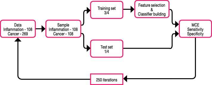
Metabolic profiles of plasma from 347 controls, 269 cancer patients and 108 patients with inflammation were obtained by H-NMR spectroscopy. Models to discriminate between groups were trained by PLS-LDA. A test set was used for independent validation. A ROC curve was built to evaluate the diagnostic performance of potential biomarkers.
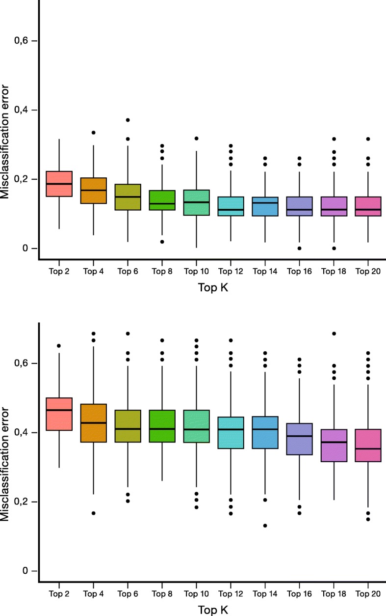
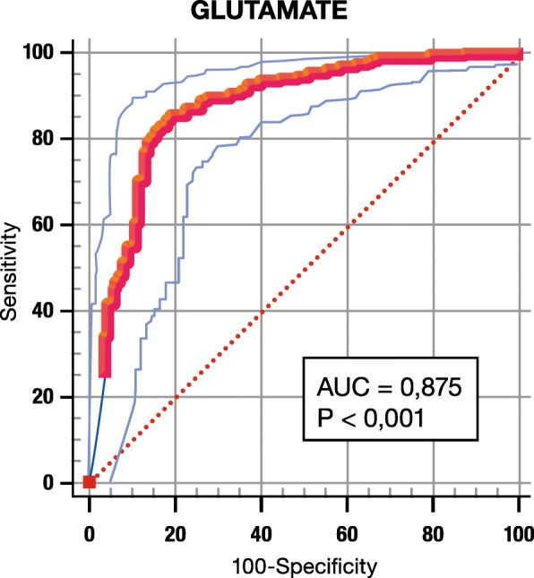
Sensitivity, specificity, PPV and NPV of PET-CT to diagnose cancer are 96, 23, 76 and 71%. Metabolic profiles differentiate between cancer and inflammation with a sensitivity of 89%, a specificity of 87% and a MCE of 12%. Removal of the glutamate metabolite results in an increase of MCE (38%) and a decrease of both sensitivity and specificity (62%), demonstrating the importance of glutamate for discrimination. At the cut-off point 0.31 on the ROC curve, the relative glutamate concentration discriminates between cancer and inflammation with a sensitivity of 85%, a specificity of 81%, and an AUC of 0.88. PPV and NPV are 92 and 69%. In PET-positive patients with a relative glutamate level ≤ 0.31 the sensitivity to diagnose cancer reaches 100% with a PPV of 94%. In PET-negative patients, a relative glutamate level > 0.31 increases the specificity of PET from 23% to 58% and results in a high NPV of 100%. In case of discrepancy between SUV and the glutamate concentration, lung cancer is missed in 19% of the cases.
This study indicates that the H-NMR-derived relative plasma concentration of glutamate allows discrimination between lung cancer and lung inflammation. A glutamate level ≤ 0.31 in PET-positive patients corresponds to the diagnosis of lung cancer with a higher specificity and PPV than PET-CT. Glutamate levels > 0.31 in patients with PET negative lung lesions is likely to correspond with inflammation. Caution is needed for patients with conflicting SUV values and glutamate concentrations. Confirmation is needed in a prospective study with external validation and by another analytical technique such as HPLC-MS.
肺部影像学常能发现可疑异常,导致需要进行补充诊断程序。本研究旨在探讨血浆的代谢指纹图谱是否可用于区分肺部炎症患者和肺癌患者。
采用 H-NMR 光谱法获取 347 名对照者、269 名癌症患者和 108 名炎症患者的血浆代谢谱。采用 PLS-LDA 对组间模型进行训练。使用测试集进行独立验证。构建 ROC 曲线以评估潜在生物标志物的诊断性能。
PET-CT 诊断癌症的敏感性、特异性、PPV 和 NPV 分别为 96%、23%、76%和 71%。代谢谱可将癌症与炎症区分开来,其敏感性为 89%,特异性为 87%,MCE 为 12%。去除谷氨酸代谢物可使 MCE 增加(38%),同时降低敏感性和特异性(62%),表明谷氨酸对鉴别诊断很重要。在 ROC 曲线的截断点 0.31 处,相对谷氨酸浓度可将癌症与炎症区分开来,其敏感性为 85%,特异性为 81%,AUC 为 0.88。PPV 和 NPV 分别为 92%和 69%。在相对谷氨酸水平≤0.31 的 PET 阳性患者中,诊断癌症的敏感性达到 100%,PPV 为 94%。在 PET 阴性患者中,相对谷氨酸水平>0.31 可将 PET 的特异性从 23%提高到 58%,并使 NPV 高达 100%。如果 SUV 值与谷氨酸浓度存在差异,则会有 19%的肺癌病例被漏诊。
本研究表明,基于 H-NMR 的相对血浆谷氨酸浓度可用于区分肺癌和肺部炎症。在 PET 阳性患者中,谷氨酸水平≤0.31 可诊断为肺癌,其特异性和 PPV 高于 PET-CT。在 PET 阴性肺部病变患者中,谷氨酸水平>0.31 可能与炎症相对应。对于 SUV 值和谷氨酸浓度存在冲突的患者需要谨慎。需要通过外部验证和其他分析技术(如 HPLC-MS)进行前瞻性研究和确认。