Department of Neurology and Neurosurgery, Montreal Neurological Institute and Hospital, McGill University, Montreal, Quebec, Canada.
McConnell Brain Imaging Centre, Montreal Neurological Institute, McGill University, Montreal, Quebec, Canada.
Ann Neurol. 2018 Oct;84(4):576-587. doi: 10.1002/ana.25324. Epub 2018 Oct 5.
To examine the relationship between carotid atherosclerosis and cerebral cortical thickness and investigate whether cortical thickness mediates the association between carotid atheroma and relative cognitive decline.
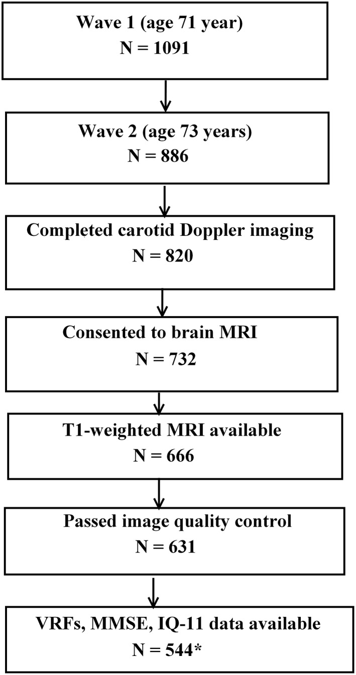
We assessed 554 community-dwelling subjects (male/female: 296/258) from the Lothian Birth Cohort 1936 who underwent brain magnetic resonance imaging and carotid Doppler ultrasound studies at age 73 years. The relationship between carotid atherosclerosis markers (internal carotid artery stenosis, intima-media thickness, velocity, pulsatility, and resistivity indexes) and vertex-wide cerebral cortical thickness was examined cross-sectionally, controlling for gender, extensive vascular risk factors (VRFs), and intelligence quotient at age 11 (IQ-11). We also determined the association between carotid stenosis and a composite measure of fluid intelligence at age 73 years. A mediation model was applied to examine whether cortical thickness mediated the relationship between carotid stenosis and cognitive function.
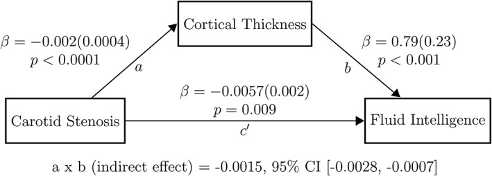
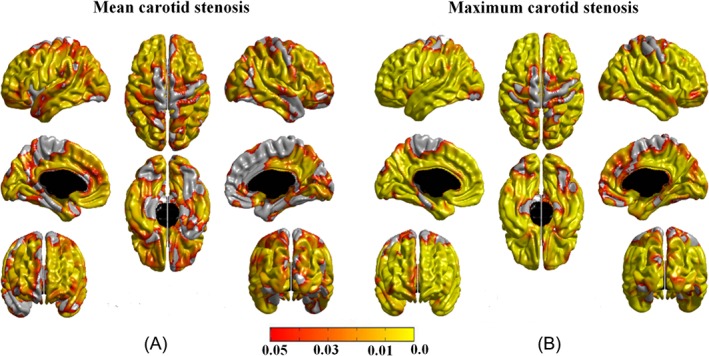
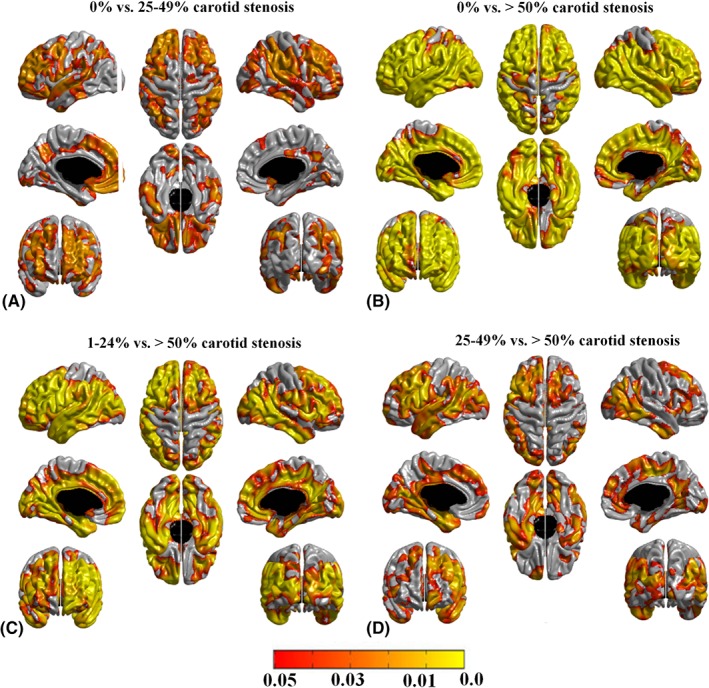
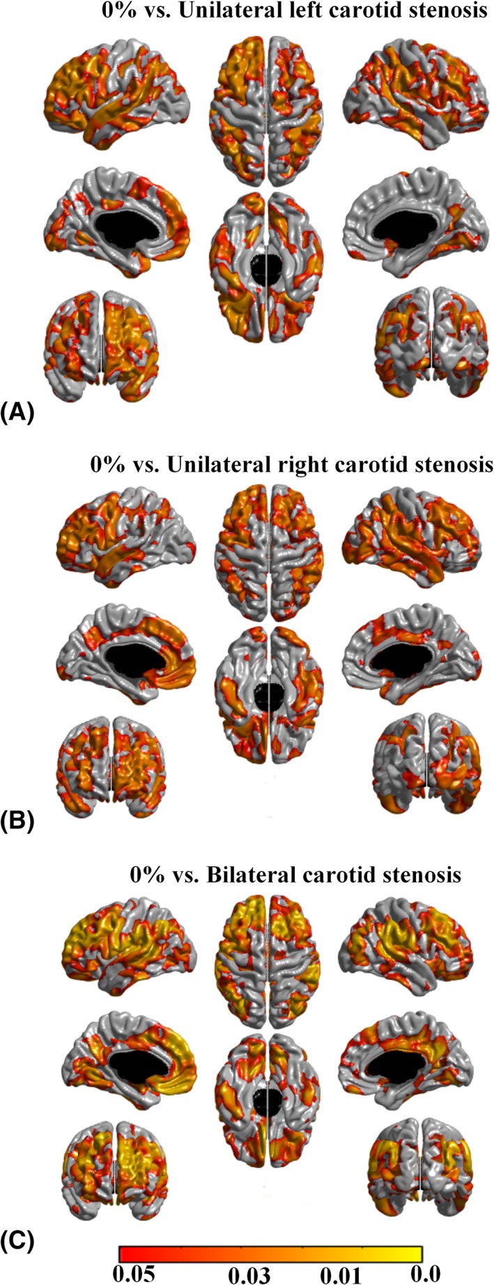
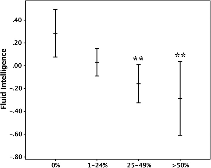
A widespread negative association was identified between carotid stenosis (median = 15%) and cerebral cortical thickness at age 73 years, independent of the side of carotid stenosis, other carotid measures, VRFs, and IQ-11. This association increased in an almost dose-response relationship from mild to severe degrees of carotid stenosis, across the anterior and posterior circulation territories. A negative association was also noted between carotid stenosis and fluid intelligence (standardized beta coefficient = -0.151, p = 0.001), which appeared partly (approximately 22%) mediated by carotid stenosis-related thinning of the cerebral cortex.
The findings suggest that carotid stenosis represents a marker of processes that accelerate aging of the cerebral cortex and cognition that is in part independent of measurable VRFs. Cortical thinning within the anterior and posterior circulation territories partially mediated the relationship between carotid atheroma and fluid intelligence. Ann Neurol 2018;84:576-587.
探讨颈动脉硬化与大脑皮质厚度之间的关系,并研究皮质厚度是否介导了颈动脉粥样硬化与相对认知衰退之间的关联。
我们评估了来自洛锡安出生队列 1936 年的 554 名居住在社区的受试者(男性/女性:296/258),他们在 73 岁时接受了大脑磁共振成像和颈动脉多普勒超声检查。我们在控制了性别、广泛的血管危险因素(VRFs)和 11 岁时的智商(IQ-11)后,检查了颈动脉硬化标志物(颈内动脉狭窄、内-中膜厚度、速度、搏动性和阻力指数)与顶点全脑皮质厚度之间的关系。我们还确定了颈内动脉狭窄与 73 岁时流体智力综合指标之间的关联。应用中介模型来检验皮质厚度是否介导了颈内动脉狭窄与认知功能之间的关系。
在控制了侧别、其他颈动脉指标、VRFs 和 IQ-11 后,在 73 岁时发现颈内动脉狭窄(中位数=15%)与大脑皮质厚度之间存在广泛的负相关关系。这种关联随着颈内动脉狭窄从轻度到重度的程度增加,在前循环和后循环区域都有增加。还观察到颈内动脉狭窄与流体智力之间存在负相关关系(标准化β系数=-0.151,p=0.001),其中部分(约 22%)是由颈动脉硬化相关的大脑皮质变薄介导的。
这些发现表明,颈内动脉狭窄代表了加速大脑皮质和认知衰老的过程的标志物,其部分独立于可测量的 VRFs。前循环和后循环区域内的皮质变薄部分介导了颈动脉粥样硬化与流体智力之间的关系。