Department of Respiratory and Critical Care Medicine, Second Affiliated Hospital of Xi'an Jiaotong University, Xi'an, China.
Department of Respiratory Medicine, Shaanxi Provincial People's Hospital, Xi'an, China.
Thorac Cancer. 2018 Nov;9(11):1366-1375. doi: 10.1111/1759-7714.12839. Epub 2018 Sep 9.
Small cell lung cancer (SCLC) is highly aggressive and is associated with a dismal prognosis. However, there are no clinically recognized biomarkers for early diagnosis. In this study, we used quantitative proteomics to build differential mitochondrial protein profiles that may be used for early diagnosis and investigated the pathogenesis of lung cancer.
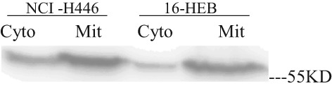
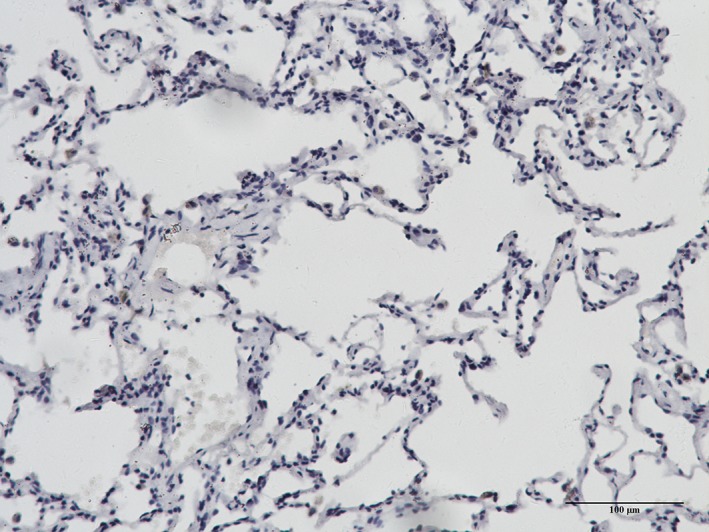
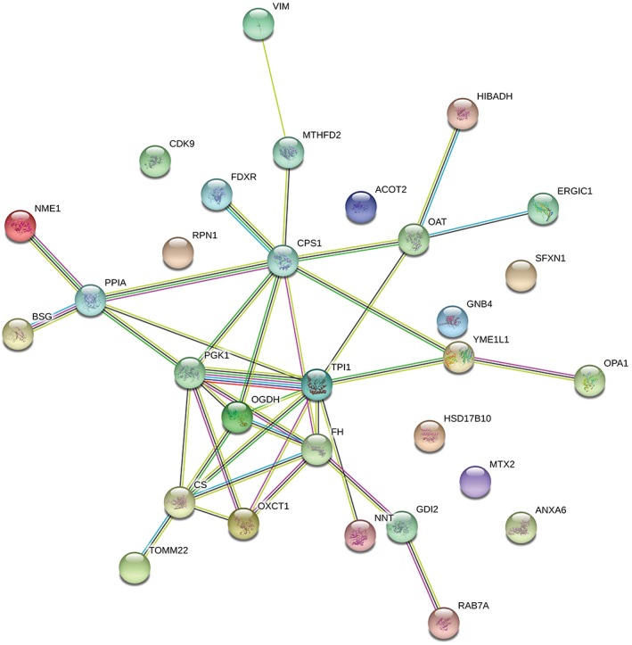
We cultured SCLC cells (NCI-H446) and normal human bronchial epithelial cells (16-HBE); mitochondria were extracted and purified using differential and Percoll density gradient centrifugation. Subsequently, we used Western blot analysis to validate mitochondrial purity and labeled proteins/peptides from NCI-H446 and 16-HBE cells using relative and absolute quantification of ectopic tags. We then analyzed mixed samples and identified proteins using two-dimensional liquid chromatography-tandem mass spectrometry. Additionally, we performed subsequent bioinformatic proteome analyses using the programs ExPASy, GOA, and STRING. Finally, the relationship between ornithine aminotransferase expression and clinicopathological features in lung cancer patients was evaluated using immunohistochemistry.
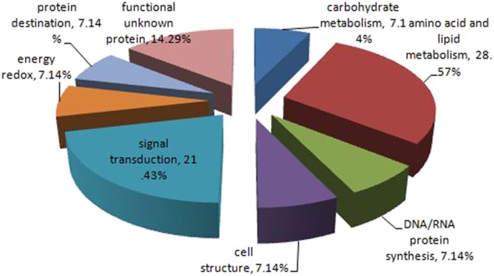
One hundred and fifty-three mitochondrial proteins were differentially expressed between 16-HBE and NCI-H446 cells. The expression of 30 proteins between 16-HBE and NCI-H446 cells increased more than 1.3-fold. The upregulation of ornithine aminotransferase was associated with pathological grade and clinical tumor node metastasis stage.
Our experiment represented a promising method for building differential mitochondrial protein profiles between NCI-H446 and 16-HBE cells. Such analysis may also help to identify novel biomarkers of lung cancer.
小细胞肺癌(SCLC)侵袭性强,预后不良。然而,目前还没有临床上公认的用于早期诊断的生物标志物。本研究采用定量蛋白质组学方法构建了可能用于早期诊断的差异线粒体蛋白质图谱,并对肺癌的发病机制进行了研究。
我们培养了小细胞肺癌细胞(NCI-H446)和正常人支气管上皮细胞(16-HBE);使用差速和 Percoll 密度梯度离心法提取和纯化线粒体。然后,我们使用 Western blot 分析来验证线粒体的纯度,并使用相对和绝对定量异位标签标记 NCI-H446 和 16-HBE 细胞的蛋白质/肽。然后,我们分析混合样品并使用二维液相色谱-串联质谱法鉴定蛋白质。此外,我们使用 ExPASy、GOA 和 STRING 程序进行了后续的生物信息学蛋白质组分析。最后,使用免疫组织化学评估鸟氨酸转氨酶表达与肺癌患者临床病理特征的关系。
16-HBE 和 NCI-H446 细胞之间有 153 种线粒体蛋白表达差异。在 16-HBE 和 NCI-H446 细胞之间,有 30 种蛋白质的表达增加了 1.3 倍以上。鸟氨酸转氨酶的上调与病理分级和临床肿瘤淋巴结转移分期有关。
我们的实验代表了一种有前途的方法,可以构建 NCI-H446 和 16-HBE 细胞之间的差异线粒体蛋白质图谱。这种分析方法也有助于识别肺癌的新型生物标志物。