de Aro Andrea Aparecida, Carneiro Giane Daniela, Teodoro Luis Felipe R, da Veiga Fernanda Cristina, Ferrucci Danilo Lopes, Simões Gustavo Ferreira, Simões Priscyla Waleska, Alvares Lúcia Elvira, de Oliveira Alexandre Leite R, Vicente Cristina Pontes, Gomes Caio Perez, Pesquero João Bosco, Esquisatto Marcelo Augusto M, de Campos Vidal Benedicto, Pimentel Edson Rosa
Department of Structural and Functional Biology, Institute of Biology, State University of Campinas⁻UNICAMP, Charles Darwin, s/n, CP 6109, 13083-970 Campinas, SP, Brazil.
Biomedical Sciences Graduate Program, Herminio Ometto University Center⁻UNIARARAS, 13607-339 Araras, SP, Brazil.
Cells. 2018 Aug 31;7(9):127. doi: 10.3390/cells7090127.
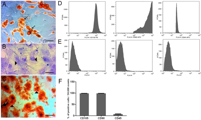
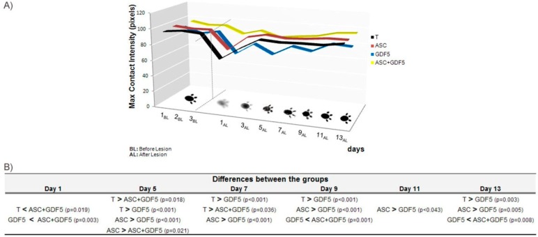
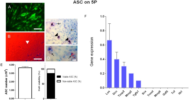
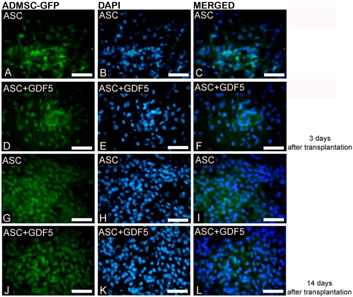
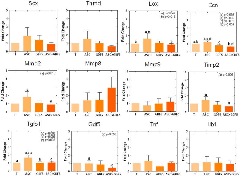
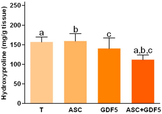
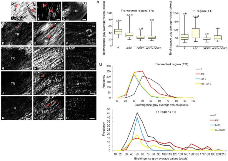
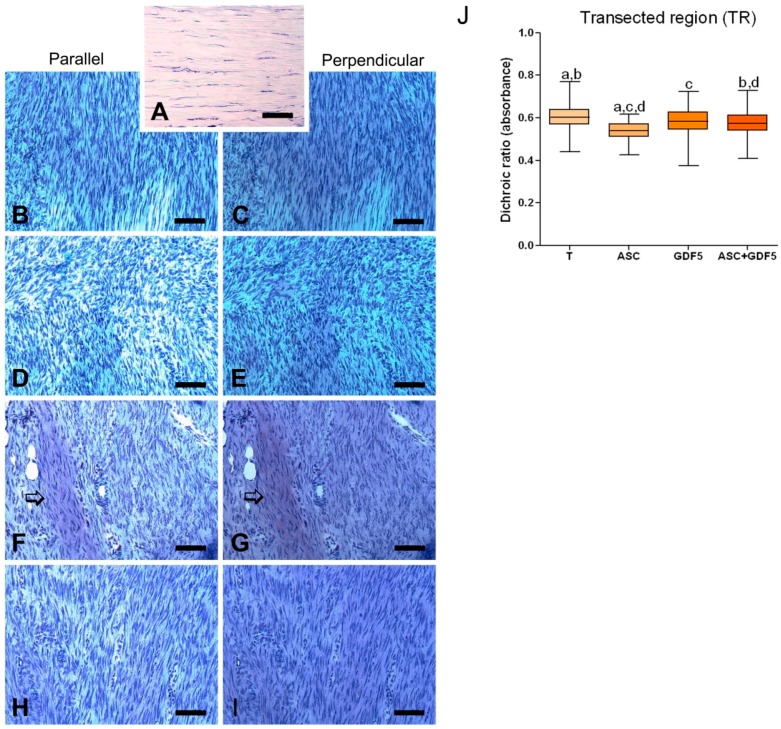
Tendon injuries represent a clinical challenge in regenerative medicine because their natural repair process is complex and inefficient. The high incidence of tendon injuries is frequently associated with sports practice, aging, tendinopathies, hypertension, diabetes mellitus, and the use of corticosteroids. The growing interest of scientists in using adipose-derived mesenchymal stem cells (ADMSC) in repair processes seems to be mostly due to their paracrine and immunomodulatory effects in stimulating specific cellular events. ADMSC activity can be influenced by GDF-5, which has been successfully used to drive tenogenic differentiation of ADMSC in vitro. Thus, we hypothesized that the application of ADMSC in isolation or in association with GDF-5 could improve Achilles tendon repair through the regulation of important remodeling genes expression. Lewis rats had tendons distributed in four groups: Transected (T), transected and treated with ADMSC (ASC) or GDF-5 (GDF5), or with both (ASC+GDF5). In the characterization of cells before application, ADMSC expressed the positive surface markers, CD90 (90%) and CD105 (95%), and the negative marker, CD45 (7%). ADMSC were also differentiated in chondrocytes, osteoblast, and adipocytes. On the 14th day after the tendon injury, GFP-ADMSC were observed in the transected region of tendons in the ASC and ASC+GDF5 groups, and exhibited and/or stimulated a similar genes expression profile when compared to the in vitro assay. ADMSC up-regulated , , and genes expression in comparison to T and ASC+GDF5 groups, which contributed to a lower proteoglycans arrangement, and to a higher collagen fiber organization and tendon biomechanics in the ASC group. The application of ADMSC in association with GDF-5 down-regulated , , , , , and genes expression, which contributed to a lower hydroxyproline concentration, lower collagen fiber organization, and to an improvement of the rats' gait 24 h after the injury. In conclusion, although the literature describes the benefic effect of GDF-5 for the tendon healing process, our results show that its application, isolated or associated with ADMSC, cannot improve the repair process of partial transected tendons, indicating the higher effectiveness of the application of ADMSC in injured Achilles tendons. Our results show that the application of ADMSC in injured Achilles tendons was more effective in relation to its association with GDF-5.
肌腱损伤是再生医学中的一项临床挑战,因为其天然修复过程复杂且效率低下。肌腱损伤的高发病率常与体育活动、衰老、肌腱病、高血压、糖尿病以及皮质类固醇的使用有关。科学家们越来越有兴趣在修复过程中使用脂肪来源的间充质干细胞(ADMSC),这似乎主要是由于它们在刺激特定细胞事件方面的旁分泌和免疫调节作用。ADMSC的活性可受生长分化因子5(GDF-5)影响,GDF-5已成功用于在体外驱动ADMSC向肌腱细胞分化。因此,我们假设单独应用ADMSC或与GDF-5联合应用可通过调节重要的重塑基因表达来改善跟腱修复。将Lewis大鼠的肌腱分为四组:横断组(T)、横断并接受ADMSC(ASC)或GDF-5(GDF5)治疗组,或两者联合治疗组(ASC+GDF5)。在应用前对细胞进行表征时,ADMSC表达阳性表面标志物CD90(90%)和CD105(95%),以及阴性标志物CD45(7%)。ADMSC还可分化为软骨细胞、成骨细胞和脂肪细胞。在肌腱损伤后第14天,在ASC和ASC+GDF5组肌腱的横断区域观察到绿色荧光蛋白标记的ADMSC(GFP-ADMSC),与体外试验相比,其表现出和/或刺激了相似的基因表达谱。与T组和ASC+GDF5组相比,ADMSC上调了 、 和 基因的表达,这导致ASC组蛋白聚糖排列减少,胶原纤维组织增加,肌腱生物力学性能提高。ADMSC与GDF-5联合应用下调了 、 、 、 、 和 基因的表达,这导致羟脯氨酸浓度降低,胶原纤维组织减少,并在损伤后24小时改善了大鼠的步态。总之,尽管文献描述了GDF-5对肌腱愈合过程的有益作用,但我们的结果表明,单独应用或与ADMSC联合应用GDF-5并不能改善部分横断肌腱的修复过程,这表明ADMSC应用于损伤的跟腱更有效。我们的结果表明,ADMSC应用于损伤的跟腱比与GDF-5联合应用更有效。