Department of Structural and Functional Biology, Institute of Biology, University of Campinas⁻UNICAMP, Charles Darwin, s/n, CP 6109, 13083-970 Campinas, SP, Brazil.
Department of Biochemistry and Tissue Biology, Institute of Biology, University of Campinas⁻UNICAMP, Charles Darwin, s/n, CP 6109, 13083-970 Campinas, SP, Brazil.
Cells. 2019 Jan 16;8(1):56. doi: 10.3390/cells8010056.
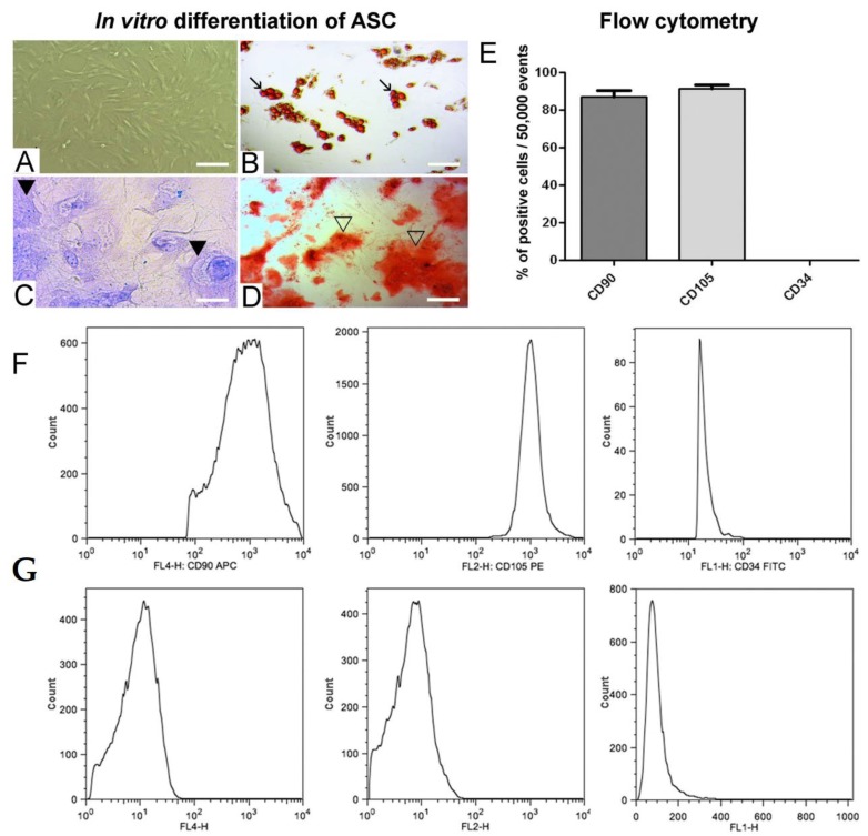
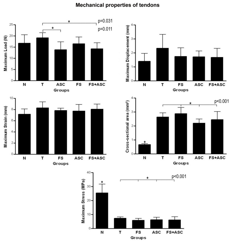
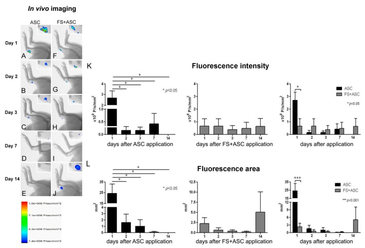
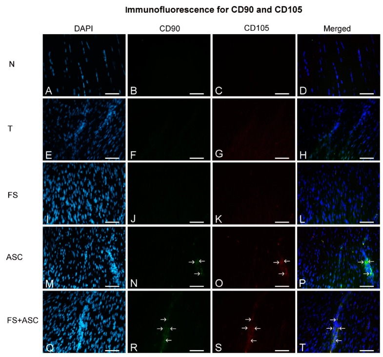
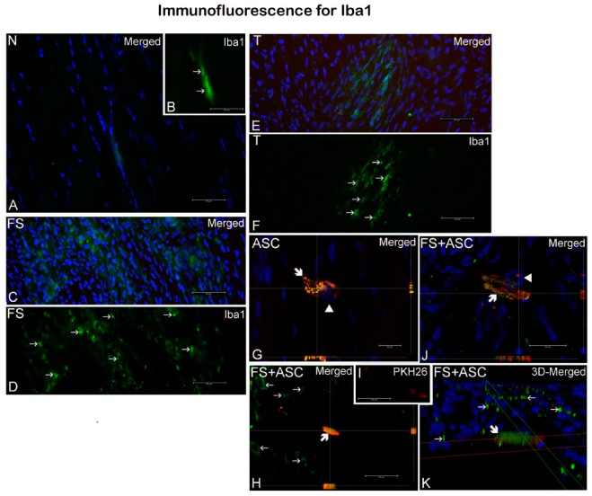
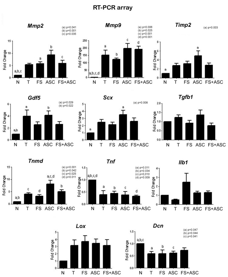
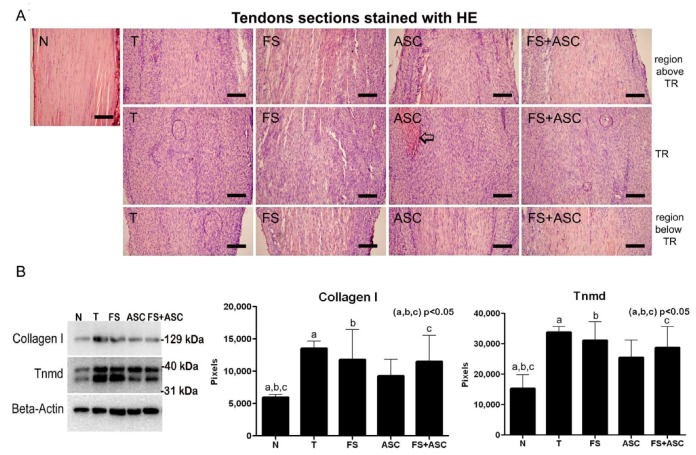
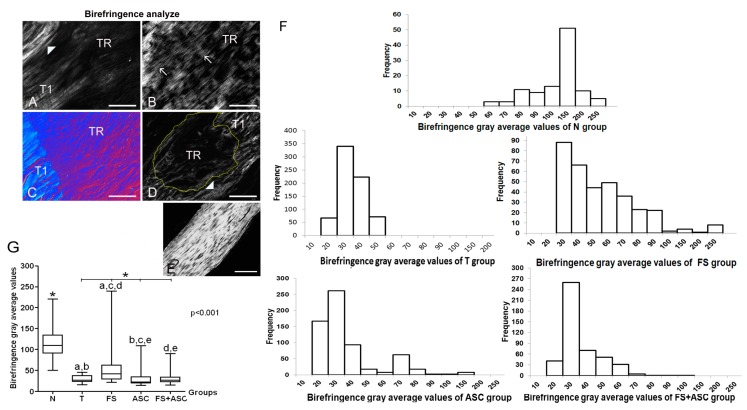
Tissue engineering and cell-based therapy combine techniques that create biocompatible materials for cell survival, which can improve tendon repair. This study seeks to use a new fibrin sealant (FS) derived from the venom of , a biodegradable three-dimensional scaffolding produced from animal components only, associated with adipose-derived stem cells (ASC) for application in tendons injuries, considered a common and serious orthopedic problem. Lewis rats had tendons distributed in five groups: normal (N), transected (T), transected and FS (FS) or ASC (ASC) or with FS and ASC (FS + ASC). The in vivo imaging showed higher quantification of transplanted PKH26-labeled ASC in tendons of FS + ASC compared to ASC on the 14th day after transection. A small number of Iba1 labeled macrophages carrying PKH26 signal, probably due to phagocytosis of dead ASC, were observed in tendons of transected groups. ASC up-regulated the gene expression in the transection region when compared to N, T and FS groups and the expression of and genes in relation to the N group. FS group presented a greater organization of collagen fibers, followed by FS + ASC and ASC in comparison to N. Tendons from ASC group presented higher hydroxyproline concentration in relation to N and the transected tendons of T, FS and FS + ASC had a higher amount of collagen I and tenomodulin in comparison to N group. Although no marked differences were observed in the other biomechanical parameters, T group had higher value of maximum load compared to the groups ASC and FS + ASC. In conclusion, the FS kept constant the number of transplanted ASC in the transected region until the 14th day after injury. Our data suggest this FS to be a good scaffold for treatment during tendon repair because it was the most effective one regarding tendon organization recovering, followed by the FS treatment associated with ASC and finally by the transplanted ASC on the 21st day. Further investigations in long-term time points of the tendon repair are needed to analyze if the higher tissue organization found with the FS scaffold will improve the biomechanics of the tendons.
组织工程和基于细胞的治疗结合了创造细胞存活的生物相容性材料的技术,这可以改善肌腱修复。本研究旨在使用一种新的纤维蛋白密封剂(FS),该密封剂源自毒液,是一种仅由动物成分制成的可生物降解的三维支架,与脂肪来源的干细胞(ASC)一起应用于肌腱损伤,这被认为是一种常见且严重的骨科问题。Lewis 大鼠的肌腱分布在五个组中:正常(N)、横断(T)、横断+FS(FS)或 ASC(ASC)或 FS+ASC。体内成像显示,在横断后第 14 天,与 ASC 相比,FS+ASC 中移植的 PKH26 标记的 ASC 数量更高。在横断组的肌腱中观察到少量携带 PKH26 信号的 Iba1 标记的巨噬细胞,可能是由于对死亡 ASC 的吞噬作用。与 N、T 和 FS 组相比,ASC 上调了横断区的 基因表达,与 N 组相比, 基因和 基因的表达。FS 组的胶原纤维组织比 N 组更有组织,FS+ASC 和 ASC 组次之。与 N 组相比,ASC 组的肌腱羟脯氨酸浓度更高,而 T、FS 和 FS+ASC 的横断肌腱的胶原 I 和肌腱调蛋白含量高于 N 组。尽管在其他生物力学参数中没有观察到明显差异,但 T 组的最大负荷值高于 ASC 和 FS+ASC 组。结论:FS 保持了横断区移植 ASC 的数量不变,直到损伤后第 14 天。我们的数据表明,这种 FS 是治疗肌腱修复的良好支架,因为它在组织恢复方面最有效,其次是 FS 治疗联合 ASC,最后是 21 天移植的 ASC。需要进行更长期的肌腱修复时间点的进一步研究,以分析 FS 支架中发现的更高组织组织是否会改善肌腱的生物力学性能。