Department of Chemistry, Oregon State University, Corvallis, OR 97331, USA.
Institute of Molecular Biology and Department of Physics, University of Oregon, Eugene, OR 97403, USA.
Molecules. 2018 Sep 1;23(9):2226. doi: 10.3390/molecules23092226.
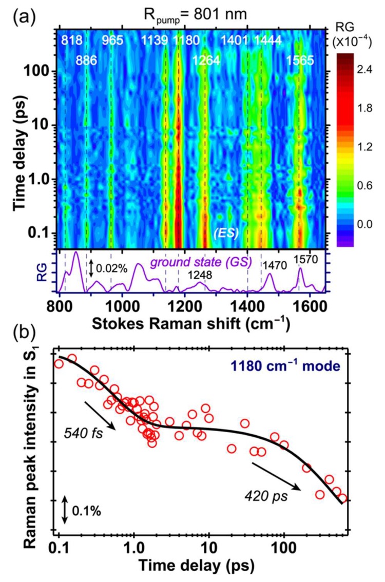
Tracking vibrational motions during a photochemical or photophysical process has gained momentum, due to its sensitivity to the progression of reaction and change of environment. In this work, we implemented an advanced ultrafast vibrational technique, femtosecond-stimulated Raman spectroscopy (FSRS), to monitor the excited state structural evolution of an engineered green fluorescent protein (GFP) single-site mutant S205V. This mutation alters the original excited state proton transfer (ESPT) chain. By strategically tuning the Raman pump to different wavelengths (i.e., 801, 539, and 504 nm) to achieve pre-resonance with transient excited state electronic bands, the characteristic Raman modes of the excited protonated (A*) chromophore species and intermediate deprotonated (I*) species can be selectively monitored. The inhomogeneous distribution/population of A* species go through ESPT with a similar ~300 ps time constant, confirming that bridging a water molecule to protein residue T203 in the ESPT chain is the rate-limiting step. Some A* species undergo vibrational cooling through high-frequency motions on the ~190 ps time scale. At early times, a portion of the largely protonated A* species could also undergo vibrational cooling or return to the ground state with a ~80 ps time constant. On the photoproduct side, a ~1330 cm delocalized motion is observed, with dispersive line shapes in both the Stokes and anti-Stokes FSRS with a pre-resonance Raman pump, which indicates strong vibronic coupling, as the mode could facilitate the I* species to reach a relatively stable state (e.g., the main fluorescent state) after conversion from A*. Our findings disentangle the contributions of various vibrational motions active during the ESPT reaction, and offer new structural dynamics insights into the fluorescence mechanisms of engineered GFPs and other analogous autofluorescent proteins.
追踪光化学或光物理过程中的振动运动由于其对反应进程和环境变化的敏感性而受到关注。在这项工作中,我们采用了先进的超快振动技术——飞秒受激拉曼光谱(FSRS)来监测工程化绿色荧光蛋白(GFP)单一位点突变体 S205V 的激发态结构演变。该突变改变了原始的激发态质子转移(ESPT)链。通过策略性地将拉曼泵调谐到不同波长(即 801nm、539nm 和 504nm),以与瞬态激发态电子带预共振,可选择性地监测激发质子化(A*)发色团物种和中间去质子化(I*)物种的特征拉曼模式。A物种的非均匀分布/种群经历 ESPT 具有相似的~300 ps 时间常数,这证实了在 ESPT 链中 bridg 一个水分子到蛋白质残基 T203 是限速步骤。一些 A物种通过在190 ps 时间尺度上的高频运动经历振动冷却。在早期,很大一部分高度质子化的 A*物种也可以通过80 ps 时间常数的振动冷却或返回基态。在光产物侧,观察到~1330 cm 的弥散运动,在具有预共振拉曼泵的斯托克斯和反斯托克斯 FSRS 中具有弥散线形状,这表明强振子耦合,因为该模式可以促进 I物种在从 A转化后达到相对稳定的状态(例如,主要荧光状态)。我们的发现解开了在 ESPT 反应过程中活跃的各种振动运动的贡献,并为工程 GFP 和其他类似自发荧光蛋白的荧光机制提供了新的结构动力学见解。