Department of Medicine, Bone & Joint Research Group, University of Auckland, Auckland, New Zealand.
Department of Rheumatology, Mater Misericordiae University Hospital, Dublin, Ireland.
Arthritis Res Ther. 2018 Sep 10;20(1):208. doi: 10.1186/s13075-018-1704-y.
Bone erosion is a frequent complication of gout and is strongly associated with tophi, which are lesions comprising inflammatory cells surrounding collections of monosodium urate (MSU) crystals. Osteocytes are important cellular mediators of bone remodeling. The aim of this study was to investigate the direct effects of MSU crystals and indirect effects of MSU crystal-induced inflammation on osteocytes.
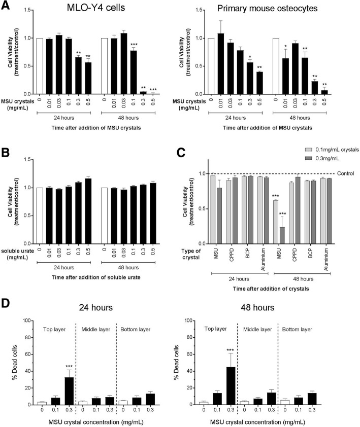
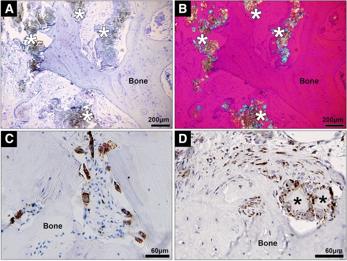
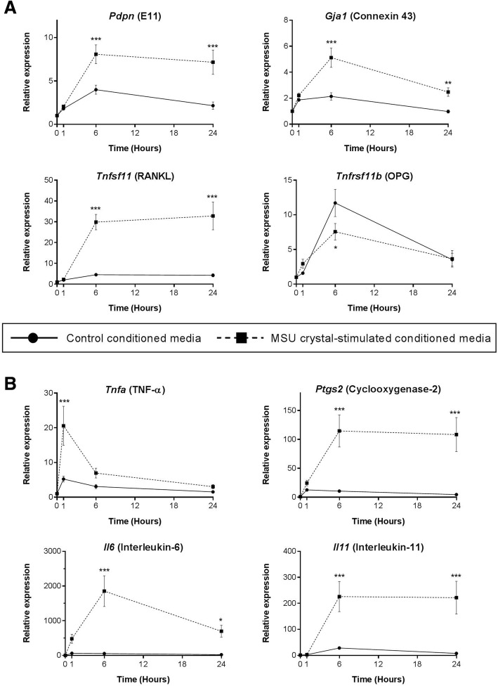
For direct assays, MSU crystals were added to MLO-Y4 osteocyte cell line cultures or primary mouse osteocyte cultures. For indirect assays, the RAW264.7 macrophage cell line was cultured with or without MSU crystals, and conditioned medium from these cultures was added to MLO-Y4 cells. MLO-Y4 cell viability was assessed using alamarBlue® and LIVE/DEAD® assays, and MLO-Y4 cell gene expression and protein expression were assessed by real-time polymerase chain reaction (PCR) and enzyme-linked immunosorbent assay (ELISA), respectively. Histological analysis was used to examine the relationship between MSU crystals, inflammatory cells, and osteocytes in human joints affected by tophaceous gout.
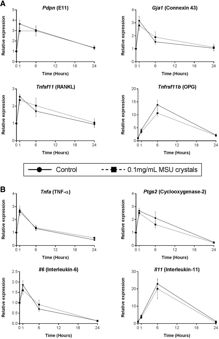
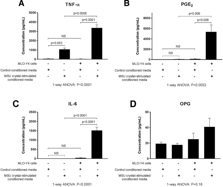
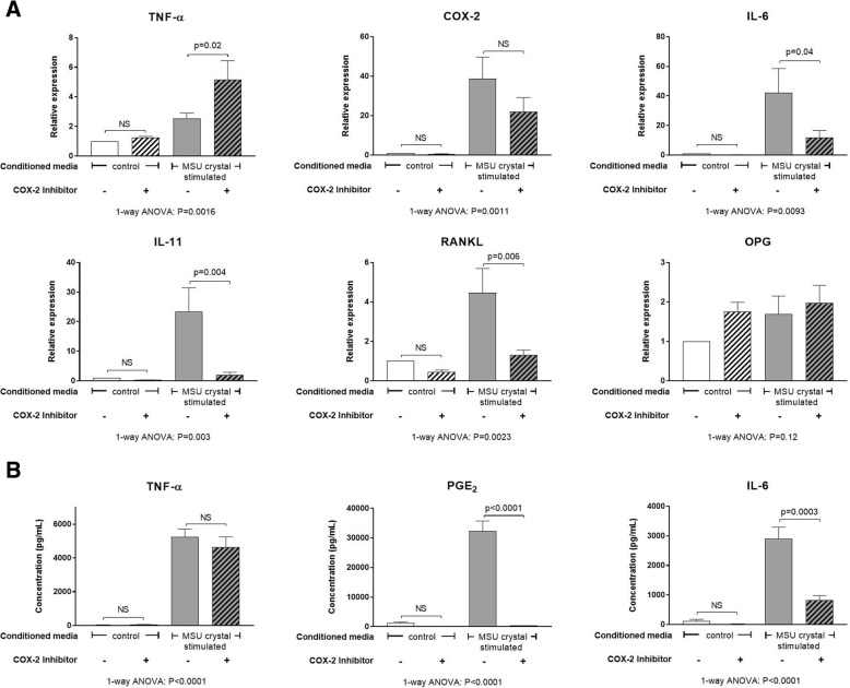
In direct assays, MSU crystals reduced MLO-Y4 cell and primary mouse osteocyte viability but did not alter MLO-Y4 cell gene expression. In contrast, conditioned medium from MSU crystal-stimulated RAW264.7 macrophages did not affect MLO-Y4 cell viability but significantly increased MLO-Y4 cell expression of osteocyte-related factors including E11, connexin 43, and RANKL, and inflammatory mediators such as interleukin (IL)-6, IL-11, tumor necrosis factor (TNF)-α and cyclooxygenase-2 (COX-2). Inhibition of COX-2 in MLO-Y4 cells significantly reduced the indirect effects of MSU crystals. In histological analysis, CD68 macrophages and MSU crystals were identified in close proximity to osteocytes within bone. COX-2 expression was also observed in tophaceous joint samples.
MSU crystals directly inhibit osteocyte viability and, through interactions with macrophages, indirectly promote a shift in osteocyte function that favors bone resorption and inflammation. These interactions may contribute to disordered bone remodeling in gout.
骨质侵蚀是痛风的常见并发症,与痛风石密切相关,痛风石是由围绕单钠尿酸盐 (MSU) 晶体的炎症细胞组成的病变。成骨细胞是骨重塑的重要细胞介质。本研究旨在探讨 MSU 晶体的直接作用和 MSU 晶体诱导的炎症的间接作用对成骨细胞的影响。
直接检测时,将 MSU 晶体加入 MLO-Y4 成骨细胞系培养物或原代小鼠成骨细胞培养物中。间接检测时,将 RAW264.7 巨噬细胞系在有无 MSU 晶体的情况下进行培养,然后将这些培养物的条件培养基加入 MLO-Y4 细胞中。通过 alamarBlue®和 LIVE/DEAD®测定评估 MLO-Y4 细胞活力,通过实时聚合酶链反应 (PCR) 和酶联免疫吸附测定 (ELISA) 分别评估 MLO-Y4 细胞基因表达和蛋白表达。组织学分析用于检查人类痛风石关节中 MSU 晶体、炎症细胞和成骨细胞之间的关系。
直接检测时,MSU 晶体降低了 MLO-Y4 细胞和原代小鼠成骨细胞的活力,但没有改变 MLO-Y4 细胞的基因表达。相比之下,来自 MSU 晶体刺激的 RAW264.7 巨噬细胞的条件培养基不影响 MLO-Y4 细胞活力,但显著增加了 MLO-Y4 细胞表达的成骨细胞相关因子,包括 E11、连接蛋白 43 和 RANKL,以及炎症介质,如白细胞介素 (IL)-6、IL-11、肿瘤坏死因子 (TNF)-α 和环氧化酶-2 (COX-2)。在 MLO-Y4 细胞中抑制 COX-2 显著降低了 MSU 晶体的间接作用。在组织学分析中,CD68 巨噬细胞和 MSU 晶体被鉴定为与骨内成骨细胞紧密相邻。在痛风石关节样本中也观察到 COX-2 表达。
MSU 晶体直接抑制成骨细胞活力,并通过与巨噬细胞相互作用间接促进成骨细胞功能的转变,有利于骨吸收和炎症。这些相互作用可能导致痛风中骨重塑紊乱。