Department of Microbiology and Immunology, Loyola University of Chicago, Stritch School of Medicine, Maywood, IL, United States.
Department of Microbiology and Immunology, College of Veterinary Medicine, Cornell University, Ithaca, NY, United States.
Virology. 2018 Dec;525:1-9. doi: 10.1016/j.virol.2018.08.022. Epub 2018 Sep 8.
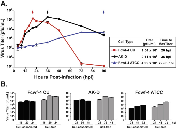
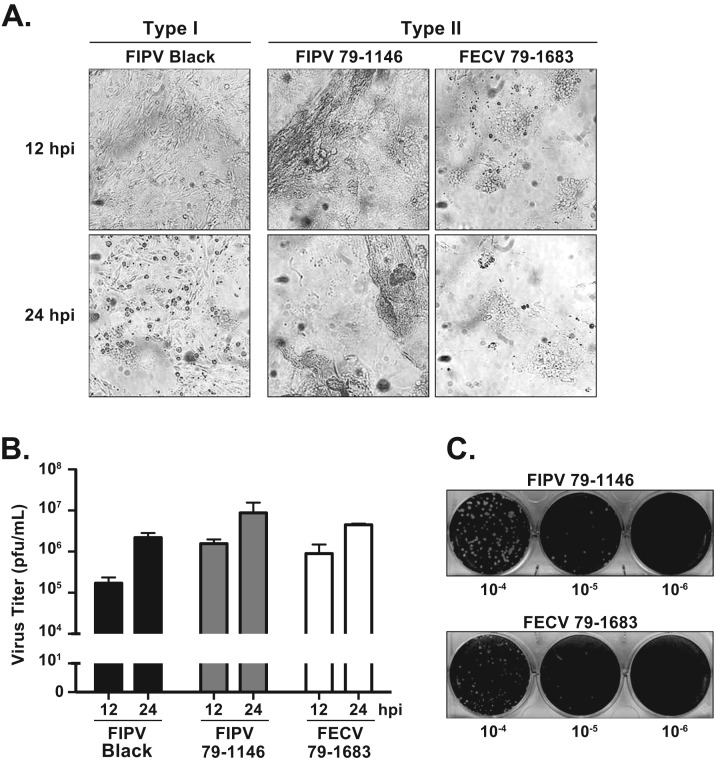
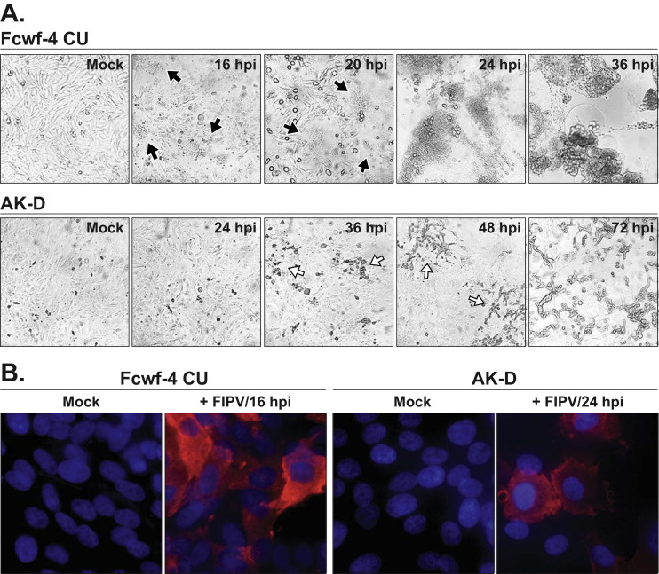
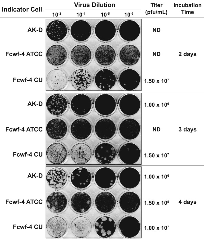
Investigating type I feline coronaviruses (FCoVs) in tissue culture is critical for understanding the basic virology, pathogenesis, and virus-host interactome of these important veterinary pathogens. This has been a perennial challenge as type I FCoV strains do not easily adapt to cell culture. Here we characterize replication kinetics and plaque formation of a model type I strain FIPV Black in Fcwf-4 cells established at Cornell University (Fcwf-4 CU). We determined that maximum virus titers (>10 pfu/mL) were recoverable from infected Fcwf-4 CU cell-free supernatant at 20 h post-infection. Type I FIPV Black and both biotypes of type II FCoV formed uniform and enumerable plaques on Fcwf-4 CU cells. Therefore, these cells were employable in a standardized plaque assay. Finally, we determined that the Fcwf-4 CU cells were morphologically distinct from feline bone marrow-derived macrophages and were less sensitive to exogenous type I interferon than were Fcwf-4 cells purchased from ATCC.
研究组织培养中的 I 型猫冠状病毒(FCoV)对于理解这些重要的兽医病原体的基础病毒学、发病机制和病毒-宿主相互作用至关重要。由于 I 型 FCoV 株不易适应细胞培养,因此这一直是一个长期存在的挑战。在这里,我们描述了在康奈尔大学(Fcwf-4 CU)建立的模型 I 型 FIPV Black 在 Fcwf-4 细胞中的复制动力学和蚀斑形成。我们确定,在感染后 20 小时,可从感染的 Fcwf-4 CU 无细胞上清液中回收最高病毒滴度(>10 pfu/mL)。I 型 FIPV Black 和两种 II 型 FCoV 生物型在 Fcwf-4 CU 细胞上形成均匀且可计数的蚀斑。因此,这些细胞可用于标准化蚀斑测定。最后,我们确定 Fcwf-4 CU 细胞在形态上与猫骨髓来源的巨噬细胞不同,并且比从 ATCC 购买的 Fcwf-4 细胞对 I 型干扰素的敏感性更低。