Kitamura Naoki, Nagami Erika, Matsushita Yumi, Kayano Tomohiko, Shibuya Izumi
Laboratory of Veterinary Physiology, Joint Department of Veterinary Medicine, Faculty of Agriculture, Tottori University, 4-101, Koyama-cho Minami, Tottori 680-8553, Japan.
IBRO Rep. 2018 Aug 18;5:33-42. doi: 10.1016/j.ibror.2018.08.002. eCollection 2018 Dec.
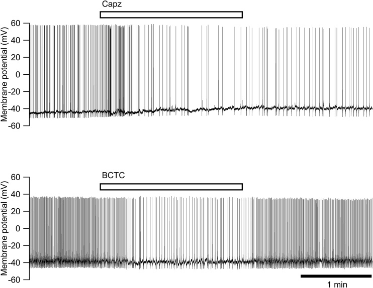
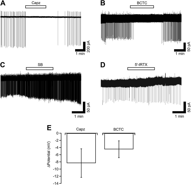
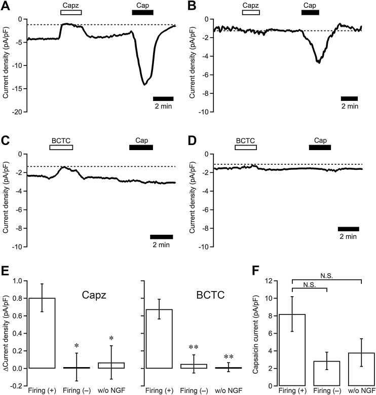
Dorsal root ganglion (DRG) neurons cultured in the presence of nerve growth factor (NGF, 100 ng/ml) often show a spontaneous action potential. Underlying mechanisms of this spontaneous firing were examined using the patch clamp technique. The spontaneous firing in the on-cell configuration was abolished by a decrease in the Na concentration and by the TRPV1 antagonists capsazepine (10 μM) and BCTC (1 μM). These responses were accompanied by hyperpolarization of the resting potential. The holding current observed in neurons voltage clamped at -60 mV in the whole-cell configuration was significantly larger in the neurons that fired spontaneously, indicating that these neurons had an additional cation conductance that caused depolarization and triggered action potentials. The holding current in the firing neurons was decreased by extracellular Na reduction, capsazepine and BCTC. The amplitudes of the capsazepine- or BCTC-sensitive component of the holding current in the spontaneously firing neurons were ten times as large as those recorded in the other neurons showing no spontaneous firing. However, the amplitudes of the current responses to capsaicin (1 μM) were not different regardless of the presence of spontaneous firing or treatment with NGF. These results indicate that chronic NGF treatment of cultured DRG neurons in rats induces a constitutively active cation conductance through TRPV1, which depolarizes the neurons and triggers spontaneous action potentials in the absence of any stimuli. Since NGF in the DRG is reported to increase after nerve injury, this NGF-mediated regulation of TRPV1 may be a cause of the pathogenesis of neuropathic pain.
在神经生长因子(NGF,100 ng/ml)存在的情况下培养的背根神经节(DRG)神经元常表现出自发动作电位。使用膜片钳技术研究了这种自发放电的潜在机制。在封接细胞模式下的自发放电可通过降低Na浓度以及使用TRPV1拮抗剂辣椒素(10 μM)和BCTC(1 μM)来消除。这些反应伴随着静息电位的超极化。在全细胞模式下钳制在 -60 mV电压的神经元中观察到的钳制电流,在自发放电的神经元中显著更大,这表明这些神经元具有额外的阳离子电导,导致去极化并触发动作电位。自发放电神经元中的钳制电流因细胞外Na减少、辣椒素和BCTC而降低。自发放电神经元中辣椒素或BCTC敏感成分的钳制电流幅度是其他无自发放电的神经元中记录到的幅度的十倍。然而,无论是否存在自发放电或用NGF处理,对辣椒素(1 μM)的电流反应幅度并无差异。这些结果表明,对大鼠培养的DRG神经元进行慢性NGF处理会通过TRPV1诱导一种组成性激活的阳离子电导,该电导使神经元去极化并在无任何刺激的情况下触发自发放电动作电位。由于据报道神经损伤后DRG中的NGF会增加,这种NGF介导的TRPV1调节可能是神经性疼痛发病机制的一个原因。