Division of Radiotherapy and Imaging, Institute of Cancer Research, London, United Kingdom; and.
Department of Nuclear Medicine, Royal Marsden NHS Foundation Trust, London, United Kingdom.
J Nucl Med. 2019 Mar;60(3):353-361. doi: 10.2967/jnumed.118.216069. Epub 2018 Sep 13.
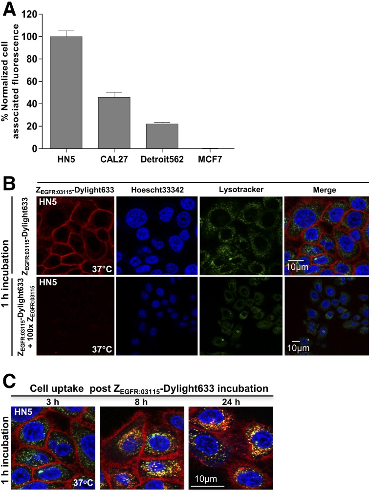
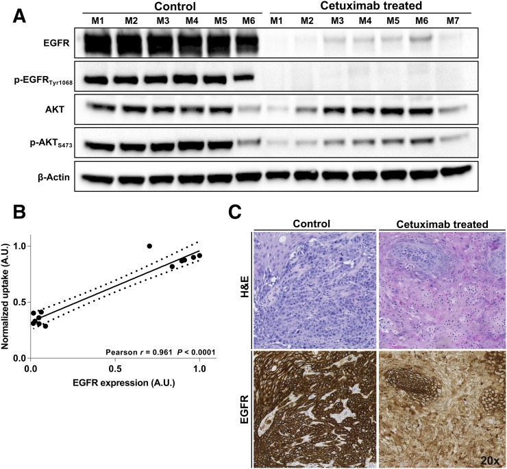
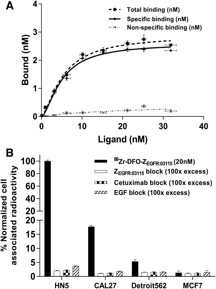
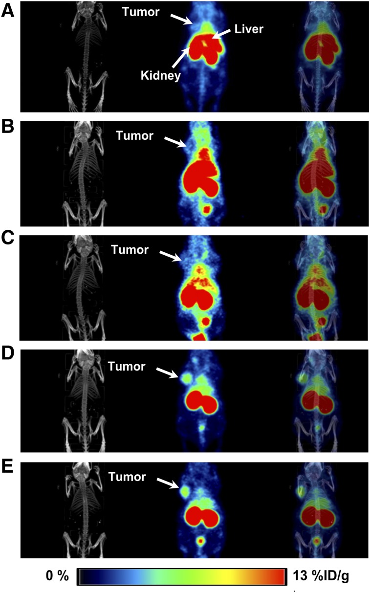
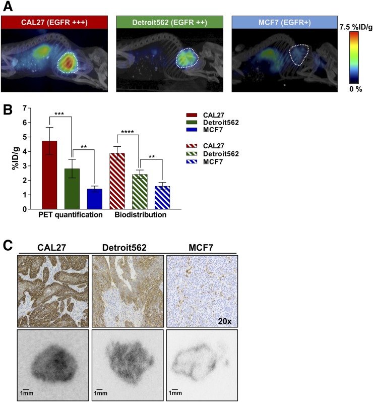
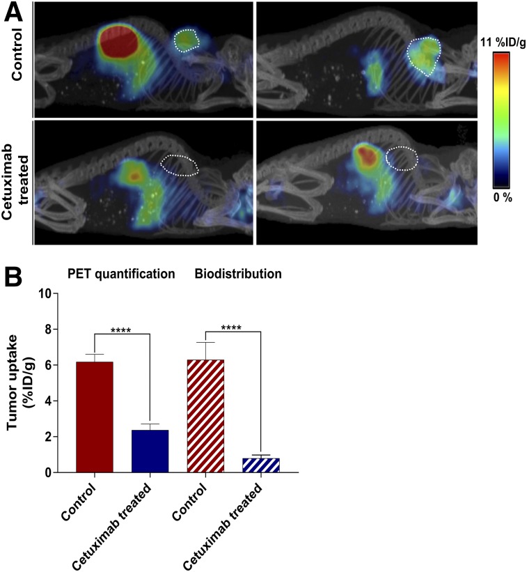
In head and neck squamous cell cancer, the human epidermal growth factor receptor 1 (EGFR) is the dominant signaling molecule among all members of the family. So far, cetuximab is the only approved anti-EGFR monoclonal antibody used for the treatment of head and neck squamous cell cancer, but despite the benefits of adding it to standard treatment regimens, attempts to define a predictive biomarker to stratify patients for cetuximab treatment have been unsuccessful. We hypothesized that imaging with EGFR-specific radioligands may facilitate noninvasive measurement of EGFR expression across the entire tumor burden and allow for dynamic monitoring of cetuximab-mediated changes in receptor expression. EGFR-specific Affibody molecule (Z) was radiolabeled with Zr and F. The radioligands were characterized in vitro and in mice bearing subcutaneous tumors with varying levels of EGFR expression. The protein dose for imaging studies was assessed by injecting Zr-deferoxamine-Z (2.4-3.6 MBq, 2 μg) either together with or 30 min after increasing amounts of unlabeled Z (1, 5, 10, 15, and 20 μg). PET images were acquired at 3, 24, and 48 h after injection, and the image quantification data were correlated with the biodistribution results. The EGFR expression and biodistribution of the tracer were assessed ex vivo by immunohistochemistry, Western blot, and autoradiography. To downregulate the EGFR level, treatment with cetuximab was performed, and F-aluminium fluoride-NOTA-Z (12 μg, 1.5-2 MBq/mouse) was used to monitor receptor changes. In vivo studies demonstrated that coinjecting 10 μg of nonlabeled molecules with Zr-deferoxamine-Z allows for clear tumor visualization 3 h after injection. The radioconjugate tumor accumulation was EGFR-specific, and PET imaging data showed a clear differentiation between xenografts with varying EGFR expression levels. A strong correlation was observed between PET analysis, ex vivo estimates of tracer concentration, and receptor expression in tumor tissues. Additionally, F-aluminium fluoride-NOTA-Z could measure receptor downregulation in response to EGFR inhibition. Z-based radioconjugates can assess different levels of EGFR level in vivo and measure receptor expression changes in response to cetuximab, indicating a potential for assessment of adequate treatment dosing with anti-EGFR antibodies.
在头颈部鳞状细胞癌中,人类表皮生长因子受体 1(EGFR)是家族中所有成员的主要信号分子。到目前为止,西妥昔单抗是唯一被批准用于治疗头颈部鳞状细胞癌的抗 EGFR 单克隆抗体,但尽管在标准治疗方案中加入它有好处,但尝试确定预测生物标志物来分层患者接受西妥昔单抗治疗一直没有成功。我们假设,使用 EGFR 特异性放射性配体进行成像可以促进对整个肿瘤负担的 EGFR 表达进行无创测量,并允许对西妥昔单抗介导的受体表达变化进行动态监测。用 Zr 和 F 对 EGFR 特异性亲和体分子(Z)进行放射性标记。在体外和表达 EGFR 水平不同的皮下肿瘤小鼠中对放射性配体进行了表征。通过注射 Zr-去铁胺-Z(2.4-3.6 MBq,2 μg),要么与未标记 Z(1、5、10、15 和 20 μg)一起,要么在 30 分钟后,评估了成像研究的蛋白质剂量。在注射后 3、24 和 48 小时采集 PET 图像,并将图像定量数据与生物分布结果相关联。通过免疫组织化学、Western blot 和放射自显影术评估了示踪剂的 EGFR 表达和生物分布。为了下调 EGFR 水平,进行了西妥昔单抗治疗,并使用 F-铝氟化物-NOTA-Z(12 μg,1.5-2 MBq/只)来监测受体变化。体内研究表明,在注射后 3 小时,与 Zr-去铁胺-Z 共注射 10 μg 的非标记分子可清楚地观察到肿瘤。放射性标记物的肿瘤积累是 EGFR 特异性的,PET 成像数据显示出不同 EGFR 表达水平的异种移植物之间的明显差异。PET 分析、肿瘤组织中外源性示踪剂浓度的体外估计值和受体表达之间观察到很强的相关性。此外,F-铝氟化物-NOTA-Z 可以测量针对 EGFR 抑制的受体下调。基于 Z 的放射性缀合物可以评估体内不同水平的 EGFR 水平,并测量针对西妥昔单抗的受体表达变化,表明有潜力评估针对 EGFR 抗体的适当治疗剂量。