Nakashima Yuko, Setou Mitsutoshi
International Mass Imaging Center and Department of Cellular and Molecular Anatomy, Hamamatsu University School of Medicine, Japan.
Preeminent Medical Photonics Education & Research Center, Japan.
Mass Spectrom (Tokyo). 2018;7(1):A0070. doi: 10.5702/massspectrometry.A0070. Epub 2018 Sep 11.
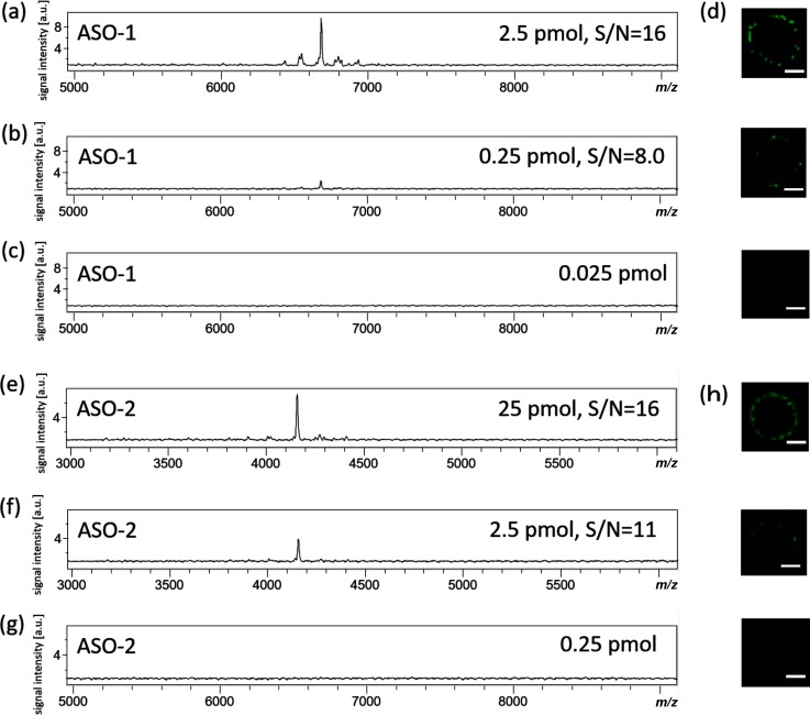
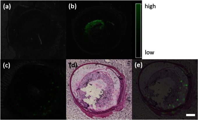
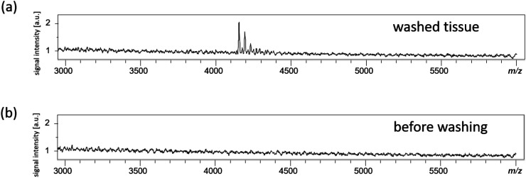
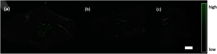
Oligonucleotide-based therapeutics such as antisense oligonucleotides, small interfering RNAs (siRNAs), decoy and aptamer have been extensively developed. To investigate the pharmacokinetics of oligonucleotide therapeutics, it is common to label a radioisotope in a nucleic acid and visualize it. However, if the labeled terminal nucleotide is decomposed by a nuclease , only the labeled nucleotide is detected, and it is impossible to observe the nucleic acid exhibiting the drug effect. The distribution of biomolecules, such as phospholipids, proteins, and glycolipids, can be obtained and visualized without labeling using matrix-assisted laser desorption/ionization imaging mass spectrometry (MALDI-IMS). MALDI-IMS is also used in pharmacokinetic analysis to visualize a parent drug and its metabolites simultaneously. In this study, we reported a methodology for oligonucleotides analysis by MALDI-IMS. When phosphorothioate antisense oligonucleotide was administered into the eyeball of rats, it reached the retina after 30 min without undergoing decomposition by nucleases.
基于寡核苷酸的治疗药物,如反义寡核苷酸、小干扰RNA(siRNA)、诱饵和适配体等已得到广泛开发。为了研究寡核苷酸治疗药物的药代动力学,通常会在核酸中标记放射性同位素并对其进行可视化。然而,如果标记的末端核苷酸被核酸酶分解,就只能检测到标记的核苷酸,而无法观察到发挥药物作用的核酸。利用基质辅助激光解吸/电离成像质谱(MALDI-IMS),无需标记就能获得并可视化生物分子(如磷脂、蛋白质和糖脂)的分布。MALDI-IMS也用于药代动力学分析,以同时可视化母体药物及其代谢物。在本研究中,我们报道了一种通过MALDI-IMS分析寡核苷酸的方法。当将硫代磷酸反义寡核苷酸注入大鼠眼球后,它在30分钟后到达视网膜,且未被核酸酶分解。