Koopmans Iris, Hendriks Djoke, Samplonius Douwe F, van Ginkel Robert J, Heskamp Sandra, Wierstra Peter J, Bremer Edwin, Helfrich Wijnand
University of Groningen, University Medical Center Groningen (UMCG), Department of Surgery, Laboratory for Translational Surgical Oncology, Groningen, The Netherlands.
Radboud University Medical Center, Department of Radiology and Nuclear Medicine, Nijmegen, The Netherlands.
Oncoimmunology. 2018 May 31;7(8):e1466016. doi: 10.1080/2162402X.2018.1466016. eCollection 2018.
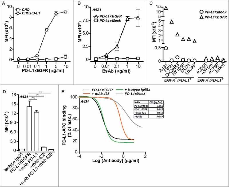
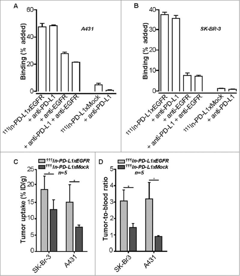
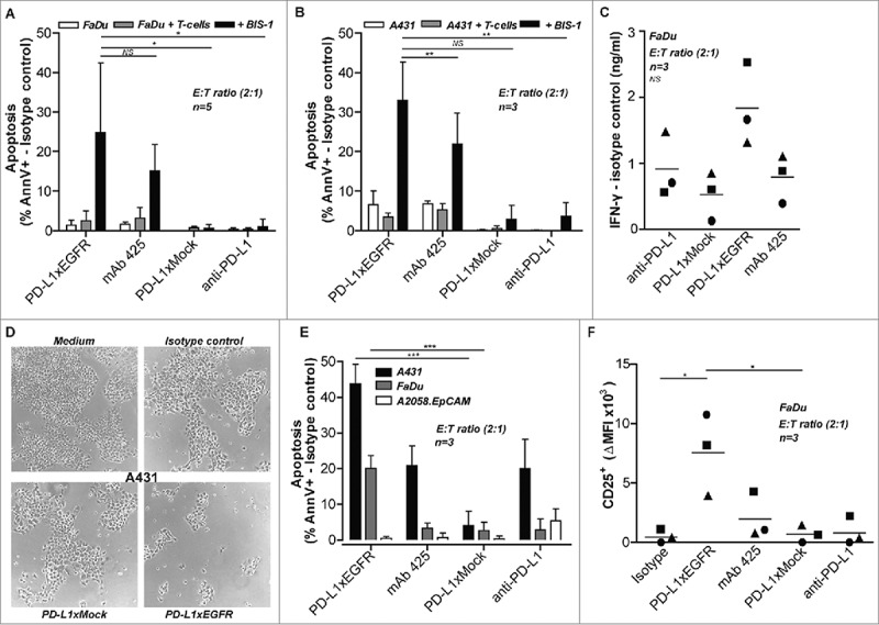
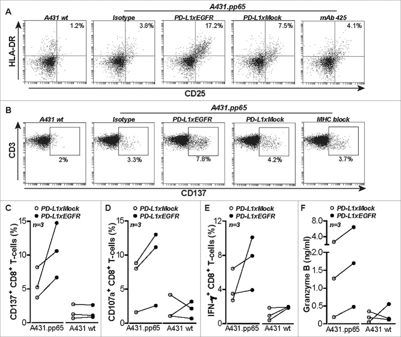
PD-L1-blocking antibodies produce significant clinical benefit in selected cancer patients by reactivating functionally-impaired antigen-experienced anticancer T cells. However, the efficacy of current PD-L1-blocking antibodies is potentially reduced by 'on-target/off-tumor' binding to PD-L1 widely expressed on normal cells. This lack of tumor selectivity may induce a generalized activation of all antigen-experienced T cells which may explain the frequent occurrence of autoimmune-related adverse events during and after treatment. To address these issues, we constructed a bispecific antibody (bsAb), designated PD-L1xEGFR, to direct PD-L1-blockade to EGFR-expressing cancer cells and to more selectively reactivate anticancer T cells. Indeed, the IC50 of PD-L1xEGFR for blocking PD-L1 on EGFR cancer cells was ∼140 fold lower compared to that of the analogous PD-L1-blocking bsAb PD-L1xMock with irrelevant target antigen specificity. Importantly, activation status, IFN-γ production, and oncolytic activity of anti-CD3xanti-EpCAM-redirected T cells was enhanced when cocultured with EGFR-expressing carcinoma cells. Similarly, the capacity of PD-L1xEGFR to promote proliferation and IFN-γ production by CMVpp65-directed CD8 effector T cells was enhanced when cocultured with EGFR-expressing CMVpp65-transfected cancer cells. In contrast, the clinically-used PD-L1-blocking antibody MEDI4736 (durvalumab) promoted T cell activation indiscriminate of EGFR expression on cancer cells. Additionally, in mice xenografted with EGFR-expressing cancer cells In-PD-L1xEGFR showed a significantly higher tumor uptake compared to In-PD-L1xMock. In conclusion, PD-L1xEGFR blocks the PD-1/PD-L1 immune checkpoint in an EGFR-directed manner, thereby promoting the selective reactivation of anticancer T cells. This novel targeted approach may be useful to enhance efficacy and safety of PD-1/PD-L1 checkpoint blockade in EGFR-overexpressing malignancies.
程序性死亡配体1(PD-L1)阻断抗体通过重新激活功能受损的抗原特异性抗癌T细胞,在部分癌症患者中产生显著的临床疗效。然而,目前的PD-L1阻断抗体的疗效可能会因与正常细胞上广泛表达的PD-L1发生“靶向脱瘤”结合而降低。这种缺乏肿瘤选择性的情况可能会导致所有抗原特异性T细胞的普遍激活,这或许可以解释治疗期间及治疗后自身免疫相关不良事件的频繁发生。为了解决这些问题,我们构建了一种双特异性抗体(bsAb),命名为PD-L1xEGFR,以将PD-L1阻断导向表达表皮生长因子受体(EGFR)的癌细胞,并更有选择性地重新激活抗癌T细胞。事实上,与具有不相关靶抗原特异性的类似PD-L1阻断双特异性抗体PD-L1xMock相比,PD-L1xEGFR阻断EGFR癌细胞上PD-L1的半数抑制浓度(IC50)低约140倍。重要的是,当与表达EGFR的癌细胞共培养时,抗CD3x抗上皮细胞黏附分子(EpCAM)重定向T细胞的激活状态、γ干扰素(IFN-γ)产生及溶瘤活性均增强。同样,当与表达EGFR的巨细胞病毒(CMV)pp65转染癌细胞共培养时,PD-L1xEGFR促进CMV pp65导向的CD8效应T细胞增殖和IFN-γ产生的能力增强。相比之下,临床使用的PD-L1阻断抗体MEDI4736(度伐鲁单抗)对癌细胞上EGFR表达情况不加区分地促进T细胞激活。此外,在接种了表达EGFR癌细胞的小鼠中,与In-PD-L1xMock相比,In-PD-L1xEGFR显示出显著更高的肿瘤摄取。总之,PD-L1xEGFR以EGFR导向的方式阻断PD-1/PD-L1免疫检查点,从而促进抗癌T细胞的选择性重新激活。这种新型靶向方法可能有助于提高EGFR过表达恶性肿瘤中PD-1/PD-L1检查点阻断的疗效和安全性。