Singh Ratnesh K, Diaz Pablo E, Binette François, Nasonkin Igor O
BioTime, Inc;
BioTime, Inc.
J Vis Exp. 2018 Aug 29(138):58274. doi: 10.3791/58274.
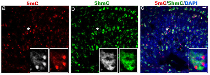
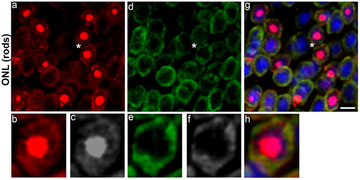
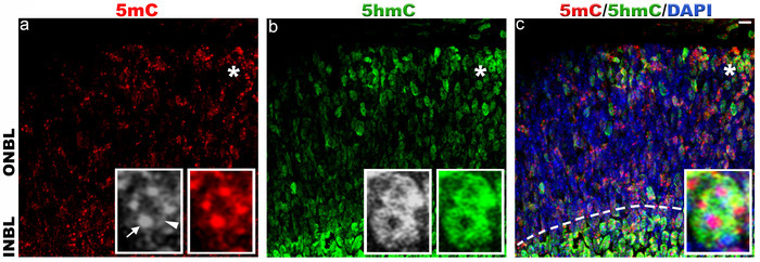
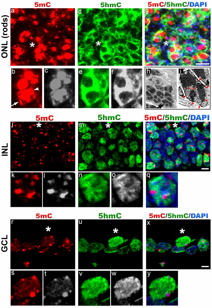
The epigenetics of retinal development is a well-studied research field, which promises to bring a new level of understanding about the mechanisms of a variety of human retinal degenerative diseases and pinpoint new treatment approaches. The nuclear architecture of mouse retina is organized in two different patterns: conventional and inverted. Conventional pattern is universal where heterochromatin is localized to the periphery of the nucleus, while active euchromatin resides in the nuclear interior. In contrast, inverted nuclear pattern is unique to the adult rod photoreceptor cell nuclei where heterochromatin localizes to the nuclear center, and euchromatin resides in the nuclear periphery. DNA methylation is predominantly observed in chromocenters. DNA methylation is a dynamic covalent modification on the cytosine residues (5-methylcytosine, 5mC) of CpG dinucleotides that are enriched in the promoter regions of many genes. Three DNA methyltransferases (DNMT1, DNMT3A and DNMT3B) participate in methylation of DNA during development. Detecting 5mC with immunohistochemical techniques is very challenging, contributing to variability in results, as all DNA bases including 5mC modified bases are hidden within the double-stranded DNA helix. However, detailed delineation of 5mC distribution during development is very informative. Here, we describe a reproducible technique for robust immunohistochemical detection of 5mC and another epigenetic DNA marker 5-hydroxymethylcytosine (5hmC), which colocalizes with the "open", transcriptionally active chromatin in developing and postmitotic mouse retina.
视网膜发育的表观遗传学是一个经过充分研究的领域,有望为多种人类视网膜退行性疾病的发病机制带来新的理解水平,并确定新的治疗方法。小鼠视网膜的核结构以两种不同模式组织:常规模式和倒置模式。常规模式普遍存在,其中异染色质定位于细胞核周边,而活跃的常染色质位于细胞核内部。相比之下,倒置核模式是成年视杆光感受器细胞核所特有的,其中异染色质定位于核中心,常染色质位于核周边。DNA甲基化主要在染色中心观察到。DNA甲基化是对富含于许多基因启动子区域的CpG二核苷酸的胞嘧啶残基(5-甲基胞嘧啶,5mC)进行的动态共价修饰。三种DNA甲基转移酶(DNMT1、DNMT3A和DNMT3B)在发育过程中参与DNA的甲基化。用免疫组织化学技术检测5mC极具挑战性,这导致结果存在差异,因为包括5mC修饰碱基在内的所有DNA碱基都隐藏在双链DNA螺旋中。然而,详细描绘发育过程中5mC的分布非常有意义。在这里,我们描述了一种可重复的技术,用于可靠地免疫组织化学检测5mC和另一种表观遗传DNA标记物5-羟甲基胞嘧啶(5hmC),5hmC在发育中和有丝分裂后的小鼠视网膜中与“开放”的、转录活跃的染色质共定位。