• 文献检索
  • 文档翻译
  • 深度研究
  • 学术资讯
  • Suppr Zotero 插件Zotero 插件
  • 邀请有礼
  • 套餐&价格
  • 历史记录
应用&插件
Suppr Zotero 插件Zotero 插件浏览器插件Mac 客户端Windows 客户端微信小程序
定价
高级版会员购买积分包购买API积分包
服务
文献检索文档翻译深度研究API 文档MCP 服务
关于我们
关于 Suppr公司介绍联系我们用户协议隐私条款
关注我们

Suppr 超能文献

核心技术专利:CN118964589B侵权必究
粤ICP备2023148730 号-1Suppr @ 2026

文献检索

告别复杂PubMed语法,用中文像聊天一样搜索,搜遍4000万医学文献。AI智能推荐,让科研检索更轻松。

立即免费搜索

文件翻译

保留排版,准确专业,支持PDF/Word/PPT等文件格式,支持 12+语言互译。

免费翻译文档

深度研究

AI帮你快速写综述,25分钟生成高质量综述,智能提取关键信息,辅助科研写作。

立即免费体验

使用首次通过门控氧-15水正电子发射断层扫描评估双心室容积和功能的可行性。

Feasibility of biventricular volume and function assessment using first-pass gated O-water PET.

作者信息

Ben Bouallègue Fayçal, Mariano-Goulart Denis, Agostini Denis, Manrique Alain

机构信息

Nuclear Medicine Department, Montpellier University Hospital, Montpellier, France.

PhyMedExp, INSERM - CNRS, Montpellier University, Montpellier, France.

出版信息

EJNMMI Res. 2018 Sep 17;8(1):92. doi: 10.1186/s13550-018-0445-x.

DOI:10.1186/s13550-018-0445-x
PMID:30225682
原文链接:https://pmc.ncbi.nlm.nih.gov/articles/PMC6141411/
Abstract

BACKGROUND

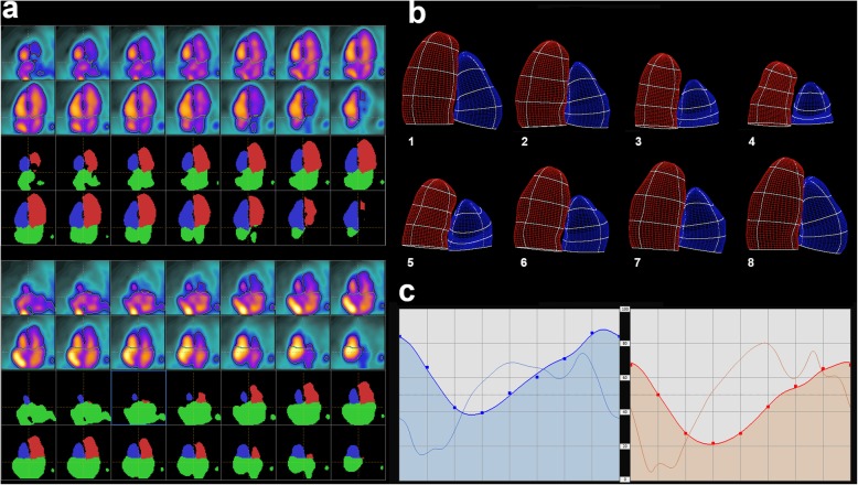
We investigated the feasibility of left ventricular (LV) and right ventricular (RV) volume and function estimation using a first-pass gated O-water PET. This prospective study included 19 patients addressed for myocardial perfusion reserve assessment using O-water PET. PET data were acquired at rest and after regadenoson stress, and gated first-pass images were reconstructed over the time range corresponding to tracer first-pass through the cardiac cavities and post-processed using TomPool software; LV and RV were segmented using a semi-automated 4D immersion algorithm. LV volumes were computed using a count-based model and a fixed threshold at 30% of the maximal activity. RV volumes were computed using a geometrical model and an adjustable threshold that was set so as to fit LV and RV stroke volumes. Ejection curves were fitted using a deformable reference curve model. LV results were compared to those obtained using Tc-sestamibi gated myocardial SPECT in terms of end-diastolic volume (EDV), end-systolic volume (ESV), stroke volume (SV), and ejection fraction (EF).

RESULTS

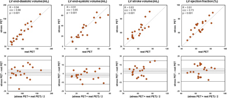
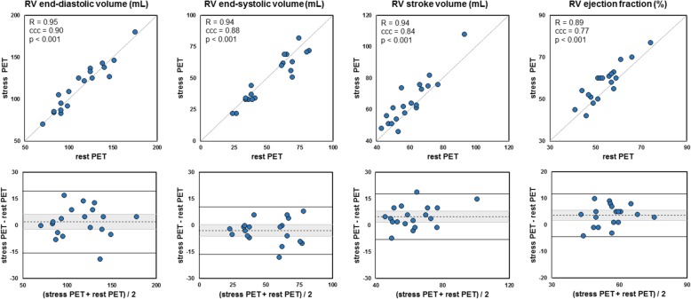
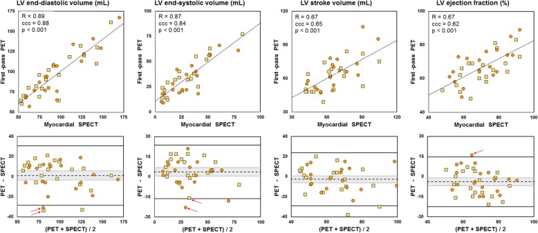
There was an excellent concordance between rest and stress PET in terms of EDV and ESV (Lin's coefficient ~ 0.85-0.90), SV (~ 0.80), and EF (~ 0.75) for both ventricles. Correlation with myocardial SPECT was high for LV EDV (Pearson's R = 0.89, p < 0.001) and ESV (R = 0.87, p < 0.001) and satisfying for LV SV (R = 0.67, p < 0.001) and EF (R = 0.67, p < 0.001). Minimal LV ESV overestimation (+ 4 mL, p = 0.03) and EF underestimation (- 4%, p = 0.01) were observed using PET.

CONCLUSIONS

Biventricular volume and function assessment are achievable using the first-pass PET, and LV parameters correlate well with those derived from gated myocardial SPECT.

摘要

背景

我们研究了使用首次通过门控氧-水正电子发射断层扫描(PET)评估左心室(LV)和右心室(RV)容积及功能的可行性。这项前瞻性研究纳入了19例因使用氧-水PET评估心肌灌注储备而接受检查的患者。PET数据在静息状态和瑞加诺生负荷后采集,门控首次通过图像在示踪剂首次通过心脏腔室的时间范围内重建,并使用TomPool软件进行后处理;使用半自动4D浸没算法对左心室和右心室进行分割。左心室容积使用基于计数的模型和最大活性30%的固定阈值进行计算。右心室容积使用几何模型和可调节阈值进行计算,该阈值设置为使左心室和右心室的每搏输出量相匹配。射血曲线使用可变形参考曲线模型进行拟合。将左心室的结果与使用锝- sestamibi门控心肌单光子发射计算机断层扫描(SPECT)获得的结果在舒张末期容积(EDV)、收缩末期容积(ESV)、每搏输出量(SV)和射血分数(EF)方面进行比较。

结果

在舒张末期容积和收缩末期容积方面,静息和负荷PET之间对于两个心室的一致性都非常好(林氏系数约为0.85 - 0.90),每搏输出量(约为0.80),射血分数(约为0.75)。左心室舒张末期容积(皮尔逊相关系数R = 0.89,p < 0.001)和收缩末期容积(R = 0.87,p < 0.001)与心肌SPECT的相关性较高,左心室每搏输出量(R = 0.67,p < 0.001)和射血分数(R = 0.67,p < 0.001)的相关性也令人满意。使用PET观察到左心室收缩末期容积有最小程度的高估(+4 mL,p = 0.03)和射血分数低估(-4%,p = 0.01)。

结论

使用首次通过PET可以实现双心室容积和功能评估,并且左心室参数与门控心肌SPECT得出的参数相关性良好。

https://cdn.ncbi.nlm.nih.gov/pmc/blobs/f600/6141411/889901f2b566/13550_2018_445_Fig4_HTML.jpg
https://cdn.ncbi.nlm.nih.gov/pmc/blobs/f600/6141411/37d68774674a/13550_2018_445_Fig1_HTML.jpg
https://cdn.ncbi.nlm.nih.gov/pmc/blobs/f600/6141411/b892e5feaa13/13550_2018_445_Fig2_HTML.jpg
https://cdn.ncbi.nlm.nih.gov/pmc/blobs/f600/6141411/91ac78627695/13550_2018_445_Fig3_HTML.jpg
https://cdn.ncbi.nlm.nih.gov/pmc/blobs/f600/6141411/889901f2b566/13550_2018_445_Fig4_HTML.jpg

相似文献

1
Feasibility of biventricular volume and function assessment using first-pass gated O-water PET.使用首次通过门控氧-15水正电子发射断层扫描评估双心室容积和功能的可行性。
EJNMMI Res. 2018 Sep 17;8(1):92. doi: 10.1186/s13550-018-0445-x.
2
Left ventricle function assessment using gated first-pass F-FDG PET: Validation against equilibrium radionuclide angiography.门控首过 F-FDG PET 评估左心室功能:与平衡放射性核素血管造影的对照验证。
J Nucl Cardiol. 2021 Apr;28(2):594-603. doi: 10.1007/s12350-019-01731-x. Epub 2019 May 1.
3
Evaluation of left and right ventricular ejection fraction and volumes from gated blood-pool SPECT in patients with dilated cardiomyopathy: comparison with cardiac MRI.门控血池 SPECT 评估扩张型心肌病患者左、右心室射血分数和容积:与心脏 MRI 的比较。
J Nucl Med. 2012 Apr;53(4):584-91. doi: 10.2967/jnumed.111.096057. Epub 2012 Mar 5.
4
Calculation of left ventricular volumes and ejection fraction from dynamic cardiac-gated O-water PET/CT: 5D-PET.基于动态心脏门控O水PET/CT(5D-PET)计算左心室容积和射血分数
EJNMMI Phys. 2017 Nov 14;4(1):26. doi: 10.1186/s40658-017-0195-2.
5
Gated F-18 FDG PET for assessment of left ventricular volumes and ejection fraction using QGS and 4D-MSPECT in patients with heart failure: a comparison with cardiac MRI.门控 F-18 FDG PET 用于评估心力衰竭患者左心室容积和射血分数:与心脏 MRI 的比较。
PLoS One. 2014 Jan 3;9(1):e80227. doi: 10.1371/journal.pone.0080227. eCollection 2014.
6
Validation of left ventricular volume measurements by gated SPECT 99mTc-labeled sestamibi imaging.通过门控单光子发射计算机断层扫描(SPECT)99mTc标记的甲氧基异丁基异腈(sestamibi)成像对左心室容积测量进行验证。
J Nucl Cardiol. 1998 Nov-Dec;5(6):574-8. doi: 10.1016/s1071-3581(98)90111-8.
7
Verification of the left ventricular ejection fraction from gated myocardial perfusion studies (GSPECT).通过门控心肌灌注显像(GSPECT)验证左心室射血分数。
Kardiol Pol. 2005 Nov;63(5):465-75; discussion 476-7.
8
Quantification of left ventricular volumes and ejection fraction from gated 99mTc-MIBI SPECT: MRI validation and comparison of the Emory Cardiac Tool Box with QGS and 4D-MSPECT.基于门控99mTc-MIBI单光子发射计算机断层扫描定量左心室容积和射血分数:磁共振成像验证及埃默里心脏工具箱与QGS和4D-MSPECT的比较
J Nucl Med. 2005 Aug;46(8):1256-63.
9
Validation of 4D-MSPECT and QGS for quantification of left ventricular volumes and ejection fraction from gated 99mTc-MIBI SPET: comparison with cardiac magnetic resonance imaging.4D-MSPECT和QGS用于门控99mTc-MIBI单光子发射计算机断层显像定量左心室容积和射血分数的验证:与心脏磁共振成像的比较
Eur J Nucl Med Mol Imaging. 2004 Apr;31(4):482-90. doi: 10.1007/s00259-003-1411-y. Epub 2004 Jan 14.
10
Influence of CT-based attenuation correction in assessment of left and right ventricular functions with count-based gated blood-pool SPECT.基于计数门控心血池 SPECT 的 CT 衰减校正对左、右心室功能评估的影响。
J Nucl Cardiol. 2011 Aug;18(4):642-9. doi: 10.1007/s12350-011-9407-7. Epub 2011 Jun 7.

引用本文的文献

1
Tales from the future-nuclear cardio-oncology, from prediction to diagnosis and monitoring.未来的核心脏肿瘤学故事——从预测到诊断和监测。
Eur Heart J Cardiovasc Imaging. 2023 Aug 23;24(9):1129-1145. doi: 10.1093/ehjci/jead168.
2
Innovative procedure for measuring left ventricular ejection fraction from F-FDG first-pass ultra-sensitive digital PET/CT images: evaluation with an anthropomorphic heart phantom.从F-FDG首次通过超灵敏数字PET/CT图像测量左心室射血分数的创新方法:使用拟人化心脏模型进行评估。
EJNMMI Phys. 2021 May 20;8(1):42. doi: 10.1186/s40658-021-00387-2.
3
Assessment of left and right ventricular functional parameters using dynamic dual-tracer [N]NH3 and [F]FDG PET/MRI.

本文引用的文献

1
First validation of myocardial flow reserve assessed by dynamic Tc-sestamibi CZT-SPECT camera: head to head comparison with O-water PET and fractional flow reserve in patients with suspected coronary artery disease. The WATERDAY study.首次评估心肌血流储备的动态 Tc-锝 CZT-SPECT 相机验证:在疑似冠心病患者中与 O-水 PET 和血流储备分数的头对头比较。WATERDAY 研究。
Eur J Nucl Med Mol Imaging. 2018 Jul;45(7):1079-1090. doi: 10.1007/s00259-018-3958-7. Epub 2018 Mar 1.
2
Effects of adenosine and regadenoson on hemodynamics measured using cardiovascular magnetic resonance imaging.腺苷和雷卡地诺生对心血管磁共振成像测量的血液动力学的影响。
J Cardiovasc Magn Reson. 2017 Dec 4;19(1):96. doi: 10.1186/s12968-017-0409-8.
3
应用动态双示踪剂 [N]NH3 和 [F]FDG PET/MRI 评估左、右心室功能参数。
J Nucl Cardiol. 2022 Jun;29(3):1003-1017. doi: 10.1007/s12350-020-02391-y. Epub 2020 Oct 22.
Calculation of left ventricular volumes and ejection fraction from dynamic cardiac-gated O-water PET/CT: 5D-PET.
基于动态心脏门控O水PET/CT(5D-PET)计算左心室容积和射血分数
EJNMMI Phys. 2017 Nov 14;4(1):26. doi: 10.1186/s40658-017-0195-2.
4
Right ventricular systolic function as a marker of prognosis after ST-elevation inferior myocardial infarction 5-year follow-up.右心室收缩功能作为ST段抬高型下壁心肌梗死后预后的标志物:5年随访
Int J Cardiol. 2016 Oct 15;221:549-53. doi: 10.1016/j.ijcard.2016.07.088. Epub 2016 Jul 5.
5
Quantitation of left ventricular ejection fraction reserve from early gated regadenoson stress Tc-99m high-efficiency SPECT.通过早期门控瑞加德松负荷 Tc-99m 高效 SPECT 定量左心室射血分数储备。
J Nucl Cardiol. 2016 Dec;23(6):1251-1261. doi: 10.1007/s12350-016-0519-y. Epub 2016 Jul 7.
6
Prognostic value of right ventricular systolic function in cardiac amyloidosis.右心室收缩功能在心脏淀粉样变性中的预后价值
Amyloid. 2016 Sep;23(3):158-167. doi: 10.1080/13506129.2016.1194264. Epub 2016 Jun 27.
7
Left Ventricular Function Assessment Using 2 Different Cadmium-Zinc-Telluride Cameras Compared with a γ-Camera with Cardiofocal Collimators: Dynamic Cardiac Phantom Study and Clinical Validation.使用2种不同碲化镉锌相机与带心脏聚焦准直器的γ相机相比评估左心室功能:动态心脏体模研究和临床验证
J Nucl Med. 2016 Sep;57(9):1370-5. doi: 10.2967/jnumed.115.168575. Epub 2016 Apr 28.
8
Measurement of LV Volumes and Function Using Oxygen-15 Water-Gated PET and Comparison With CMR Imaging.使用氧-15水门控正电子发射断层扫描测量左心室容积和功能并与心脏磁共振成像比较
JACC Cardiovasc Imaging. 2016 Dec;9(12):1472-1474. doi: 10.1016/j.jcmg.2016.01.014. Epub 2016 Apr 13.
9
The reproducibility and prognostic value of serial measurements of heart rate response to regadenoson during myocardial perfusion imaging.心肌灌注成像期间对雷加曲班心率反应进行系列测量的可重复性及预后价值
Eur J Nucl Med Mol Imaging. 2016 Jul;43(8):1493-502. doi: 10.1007/s00259-016-3380-y. Epub 2016 Apr 15.
10
Cardiac Imaging to Evaluate Left Ventricular Diastolic Function.心脏影像学评估左心室舒张功能。
JACC Cardiovasc Imaging. 2015 Sep;8(9):1071-1093. doi: 10.1016/j.jcmg.2015.07.004.