Center for In Vivo Microscopy, Department of Radiology, Duke University Medical Center, Durham, NC, USA.
Department of Radiology, School of Medicine, Duke University, Durham, NC, USA.
Brain Struct Funct. 2018 Dec;223(9):4323-4335. doi: 10.1007/s00429-018-1750-x. Epub 2018 Sep 17.
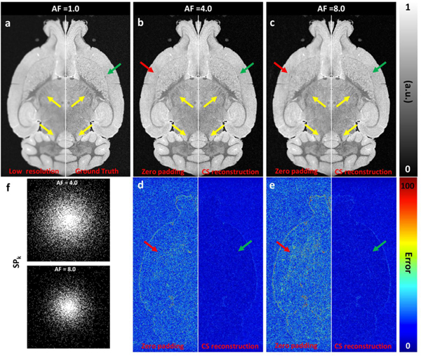
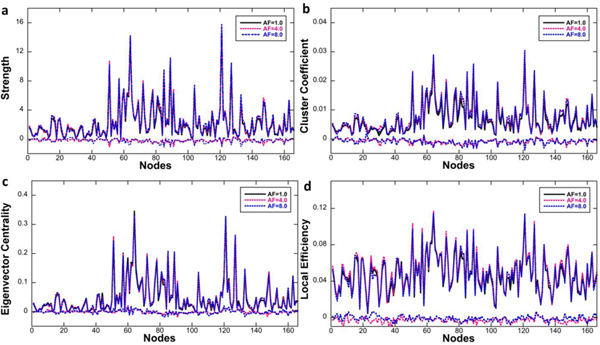
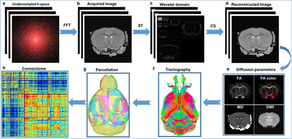
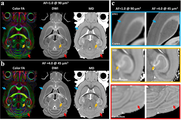
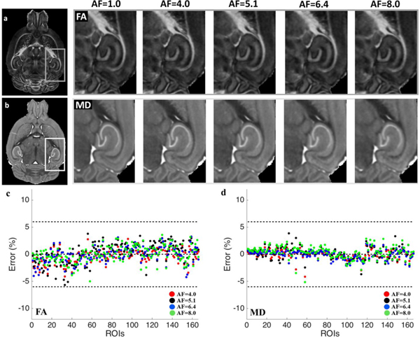
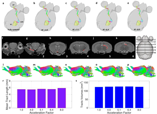
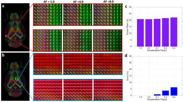
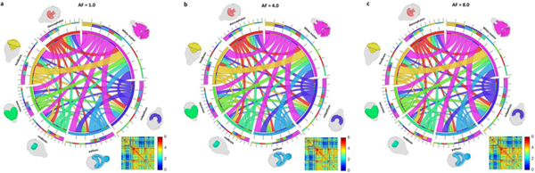
Diffusion tensor histology holds great promise for quantitative characterization of structural connectivity in mouse models of neurological and psychiatric conditions. There has been extensive study in both the clinical and preclinical domains on the complex tradeoffs between the spatial resolution, the number of samples in diffusion q-space, scan time, and the reliability of the resultant data. We describe here a method for accelerating the acquisition of diffusion MRI data to support quantitative connectivity measurements in the whole mouse brain using compressed sensing (CS). The use of CS allows substantial increase in spatial resolution and/or reduction in scan time. Compared to the fully sampled results at the same scan time, the subtle anatomical details of the brain, such as cortical layers, dentate gyrus, and cerebellum, were better visualized using CS due to the higher spatial resolution. Compared to the fully sampled results at the same spatial resolution, the scalar diffusion metrics, including fractional anisotropy (FA) and mean diffusivity (MD), showed consistently low error across the whole brain (< 6.0%) even with 8.0 times acceleration. The node properties of connectivity (strength, cluster coefficient, eigenvector centrality, and local efficiency) demonstrated correlation of better than 95.0% between accelerated and fully sampled connectomes. The acceleration will enable routine application of this technology to a wide range of mouse models of neurologic diseases.
弥散张量组织学有望对神经和精神疾病的小鼠模型的结构连接进行定量描述。在临床和临床前领域,已经对空间分辨率、扩散 q 空间中的样本数量、扫描时间以及所得数据的可靠性之间的复杂权衡进行了广泛的研究。我们在这里描述了一种使用压缩感知 (CS) 加速扩散 MRI 数据采集的方法,以支持使用全脑定量连接测量。CS 的使用可以大大提高空间分辨率和/或减少扫描时间。与在相同扫描时间下的完全采样结果相比,由于更高的空间分辨率,使用 CS 可以更好地显示大脑的细微解剖细节,如皮质层、齿状回和小脑。与在相同空间分辨率下的完全采样结果相比,标量扩散指标,包括各向异性分数 (FA) 和平均扩散系数 (MD),即使在 8 倍加速的情况下,整个大脑的误差也始终保持较低(<6.0%)。连接的节点属性(强度、聚类系数、特征向量中心度和局部效率)在加速和完全采样连接图之间表现出超过 95.0%的相关性。加速将使这项技术能够常规应用于广泛的神经疾病小鼠模型。