Department of Medicine (Cardiovascular Division), University of Virginia Health System, 1215 Lee Street, PO Box 800158, Charlottesville, VA, 22908, USA.
Department of Radiology, University of Virginia Health System, Charlottesville, VA, USA.
Curr Cardiol Rep. 2018 Sep 26;20(11):119. doi: 10.1007/s11886-018-1057-9.
This article will review the current techniques in cardiac magnetic resonance imaging (CMR) for diagnosing and assessing primary valvular heart disease.
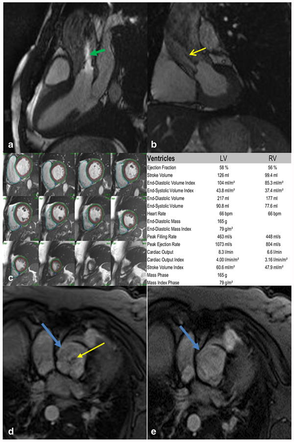
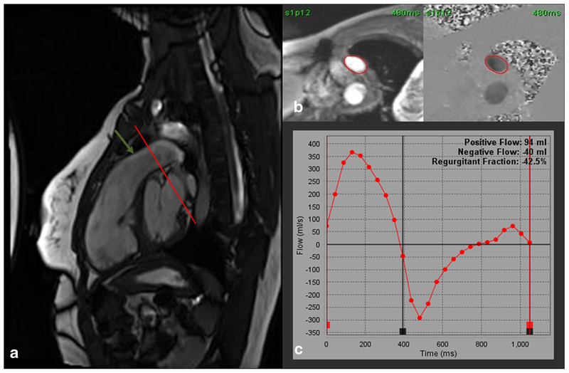
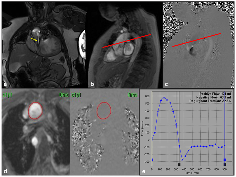
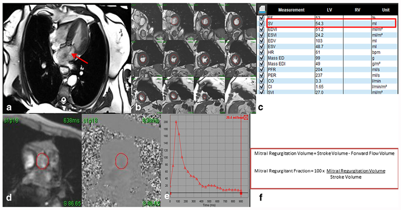
The recent advancements in CMR have led to an increased role of this modality for qualifying and quantifying various native valve diseases. Phase-contrast velocity encoded imaging is a well-established technique that can be used to quantify aortic and pulmonic flow. This technique, combined with the improved ability for CMR to obtain accurate left and right ventricular volumetrics, has allowed for increased accuracy and reproducibility in assessing valvular dysfunction. Advancements in CMR technology also allows for improved spatial and temporal resolution imaging of various valves and their regurgitant or stenotic jets. Therefore, CMR can be a powerful tool in evaluation of native valvular heart disease. The role of CMR in assessing valvular heart disease is growing and being recognized in recent guidelines. CMR has the ability to assess valve morphology along with qualifying and quantifying valvular disease. In addition, the ability to obtain accurate volumetric measurements may improve more precise management strategies and may lead to improvements in mortality and morbidity.
本文将综述心脏磁共振成像(CMR)在诊断和评估原发性心脏瓣膜病中的当前技术。
CMR 的最新进展使得该方法在定性和定量各种原生瓣膜疾病方面的作用增加。相位对比速度编码成像是一种成熟的技术,可用于量化主动脉瓣和肺动脉瓣的血流。该技术与 CMR 获得准确的左、右心室容积的能力的提高相结合,使得评估瓣膜功能障碍的准确性和可重复性提高。CMR 技术的进步还允许对各种瓣膜及其反流或狭窄射流进行更好的空间和时间分辨率成像。因此,CMR 可以成为评估原发性心脏瓣膜病的有力工具。CMR 在评估心脏瓣膜病中的作用在最近的指南中不断发展和得到认可。CMR 有能力评估瓣膜形态,同时定性和定量瓣膜疾病。此外,获得准确的容积测量值的能力可能会改善更精确的管理策略,并可能导致死亡率和发病率的降低。