MRC Laboratory of Molecular Biology, Cambridge, CB2 0QH, UK.
Department of Pathology and Laboratory Medicine, Indiana University School of Medicine, Indianapolis, IN, 46202, USA.
Acta Neuropathol. 2018 Nov;136(5):699-708. doi: 10.1007/s00401-018-1914-z. Epub 2018 Oct 1.
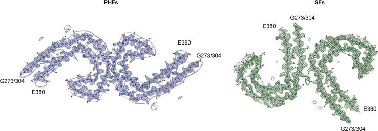
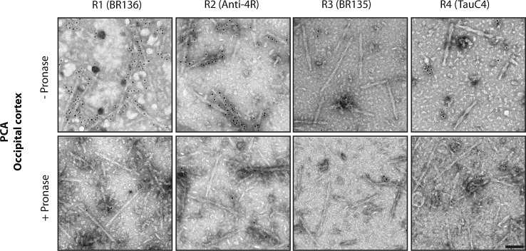
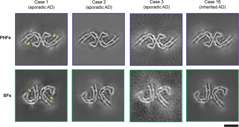
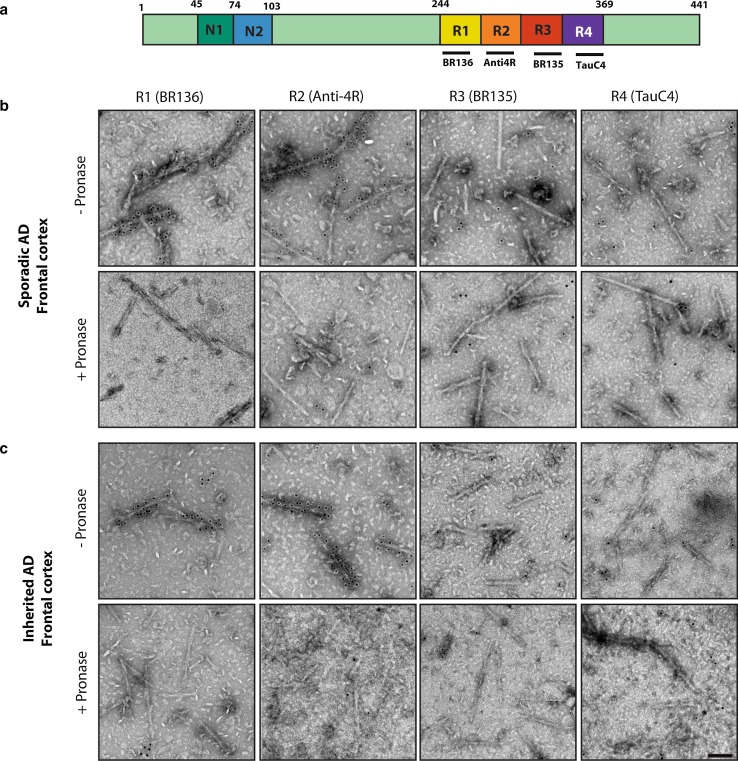
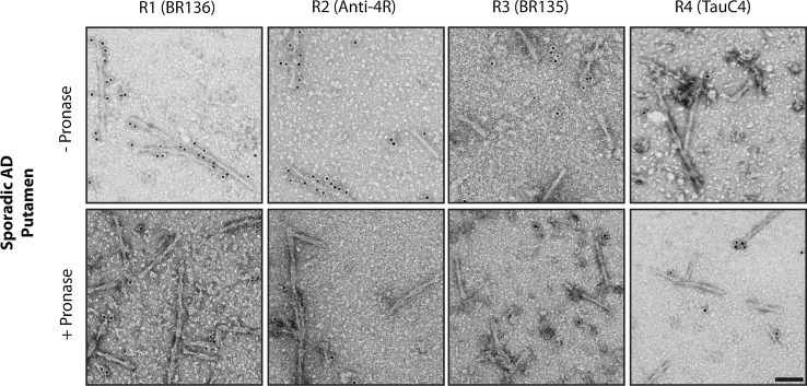
The ordered assembly of tau protein into abnormal filaments is a defining characteristic of Alzheimer's disease (AD) and other neurodegenerative disorders. It is not known if the structures of tau filaments vary within, or between, the brains of individuals with AD. We used a combination of electron cryo-microscopy (cryo-EM) and immuno-gold negative-stain electron microscopy (immuno-EM) to determine the structures of paired helical filaments (PHFs) and straight filaments (SFs) from the frontal cortex of 17 cases of AD (15 sporadic and 2 inherited) and 2 cases of atypical AD (posterior cortical atrophy). The high-resolution structures of PHFs and SFs from the frontal cortex of 3 cases of AD, 2 sporadic and 1 inherited, were determined by cryo-EM. We also used immuno-EM to study the PHFs and SFs from a number of cortical and subcortical brain regions. PHFs outnumbered SFs in all AD cases. By cryo-EM, PHFs and SFs were made of two C-shaped protofilaments with a combined cross-β/β-helix structure, as described previously for one case of AD. The higher resolution structures obtained here showed two additional amino acids at each end of the protofilament. The immuno-EM findings, which indicated the presence of repeats 3 and 4, but not of the N-terminal regions of repeats 1 and 2, of tau in the filament cores of all AD cases, were consistent with the cryo-EM results. These findings show that there is no significant variation in tau filament structures between individuals with AD. This knowledge will be crucial for understanding the mechanisms that underlie tau filament formation and for developing novel diagnostics and therapies.
tau 蛋白有序组装成异常纤维是阿尔茨海默病(AD)和其他神经退行性疾病的一个特征。目前尚不清楚 AD 患者大脑内或大脑间 tau 纤维的结构是否存在差异。我们采用电子 cryo-EM(cryo-EM)和免疫胶体金负染电镜(immuno-EM)相结合的方法,对来自 17 例 AD(15 例散发性和 2 例遗传性)和 2 例非典型 AD(后部皮质萎缩)患者额叶的成对螺旋丝(PHF)和直丝(SF)的结构进行了测定。通过 cryo-EM 确定了 3 例 AD(2 例散发性和 1 例遗传性)患者额叶 PHF 和 SF 的高分辨率结构。我们还使用 immuno-EM 研究了来自多个皮质和皮质下脑区的 PHF 和 SF。在所有 AD 病例中,PHF 的数量均多于 SF。通过 cryo-EM,PHF 和 SF 由两个 C 形原纤维组成,具有结合的 cross-β/β-螺旋结构,如先前对 1 例 AD 的描述。此处获得的更高分辨率结构显示,原纤维的每个末端都有两个额外的氨基酸。免疫电镜研究表明,所有 AD 病例的纤维核心中都存在 tau 的重复 3 和 4,但不存在重复 1 和 2 的 N 端区域,这与 cryo-EM 结果一致。这些发现表明,AD 患者之间 tau 纤维结构没有明显差异。这一知识对于理解 tau 纤维形成的机制以及开发新的诊断和治疗方法至关重要。