Department of Anesthesiology, Huai'an Maternity and Child Healthcare Hospital, Yangzhou University Medical School, Huai'an, China.
The Fourth School of Clinical Medicine, The Affiliated Eye Hospital, Nanjing Medical University, Nanjing 210029, China.
Theranostics. 2018 Sep 9;8(17):4695-4709. doi: 10.7150/thno.26203. eCollection 2018.
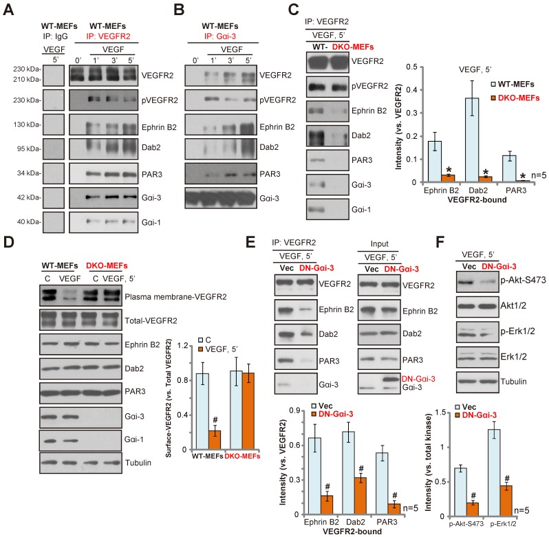
VEGF binding to VEGFR2 leads to VEGFR2 endocytosis and downstream signaling activation to promote angiogenesis. Using genetic strategies, we tested the requirement of α subunits of heterotrimeric G proteins (Gαi1/3) in the process. Gαi1/3 are located in the VEGFR2 endocytosis complex (VEGFR2-Ephrin-B2-Dab2-PAR-3), where they are required for VEGFR2 endocytosis and downstream signaling transduction. Gαi1/3 knockdown, knockout or dominant negative mutation inhibited VEGF-induced VEGFR2 endocytosis, and downstream Akt-mTOR and Erk-MAPK activation. Functional studies show that Gαi1/3 shRNA inhibited VEGF-induced proliferation, invasion, migration and vessel-like tube formation of HUVECs. , Gαi1/3 shRNA lentivirus inhibited alkali burn-induced neovascularization in mouse cornea. Further, oxygen-induced retinopathy (OIR)-induced retinal neovascularization was inhibited by intravitreal injection of Gαi1/3 shRNA lentivirus. Moreover, angiogenesis by alkali burn and OIR was significantly attenuated in Gαi1/3 double knockout mice. Significantly, Gαi1/3 proteins are upregulated in proliferative retinal tissues of proliferative diabetic retinopathy (PDR) patients. These results provide mechanistic insights into the critical role played by Gαi1/3 proteins in VEGF-induced VEGFR2 endocytosis, signaling and angiogenesis.
VEGF 与 VEGFR2 的结合导致 VEGFR2 内吞作用和下游信号激活,从而促进血管生成。我们使用遗传策略测试了异三聚体 G 蛋白(Gαi1/3)的α亚基在该过程中的需求。Gαi1/3 位于 VEGFR2 内吞复合物(VEGFR2-Ephrin-B2-Dab2-PAR-3)中,在该复合物中,它们是 VEGFR2 内吞作用和下游信号转导所必需的。Gαi1/3 的敲低、敲除或显性负突变抑制了 VEGF 诱导的 VEGFR2 内吞作用,以及下游 Akt-mTOR 和 Erk-MAPK 的激活。功能研究表明,Gαi1/3 shRNA 抑制了 VEGF 诱导的 HUVECs 的增殖、侵袭、迁移和管状结构形成。Gαi1/3 shRNA 慢病毒抑制了小鼠角膜碱烧伤诱导的新生血管形成。此外,玻璃体内注射 Gαi1/3 shRNA 慢病毒抑制了氧诱导的视网膜病变(OIR)诱导的视网膜新生血管形成。此外,在 Gαi1/3 双敲除小鼠中,碱烧伤和 OIR 引起的血管生成明显减弱。重要的是,在增生性糖尿病视网膜病变(PDR)患者的增生性视网膜组织中,Gαi1/3 蛋白上调。这些结果为 Gαi1/3 蛋白在 VEGF 诱导的 VEGFR2 内吞作用、信号转导和血管生成中的关键作用提供了机制上的见解。