Department of Otolaryngology, University of Pittsburgh, Pittsburgh, Pennsylvania.
University of Pittsburgh Hillman Cancer Center, Pittsburgh, Pennsylvania.
Cancer Immunol Res. 2018 Dec;6(12):1548-1560. doi: 10.1158/2326-6066.CIR-18-0062. Epub 2018 Oct 3.
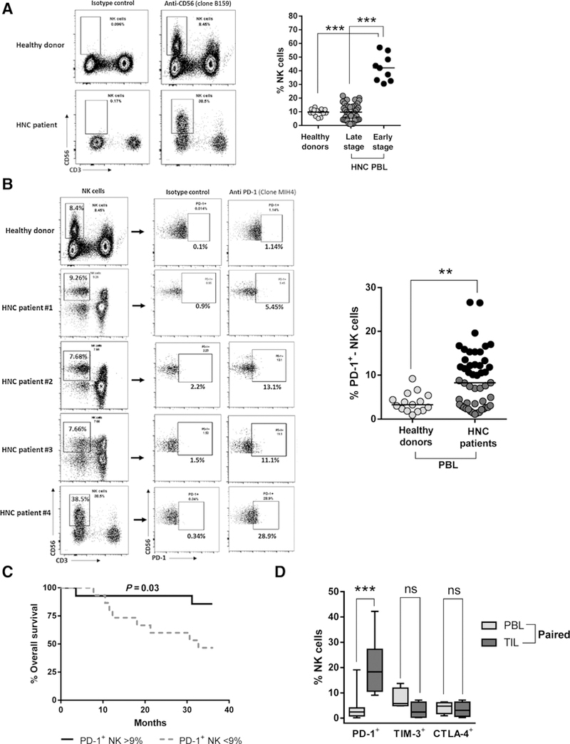
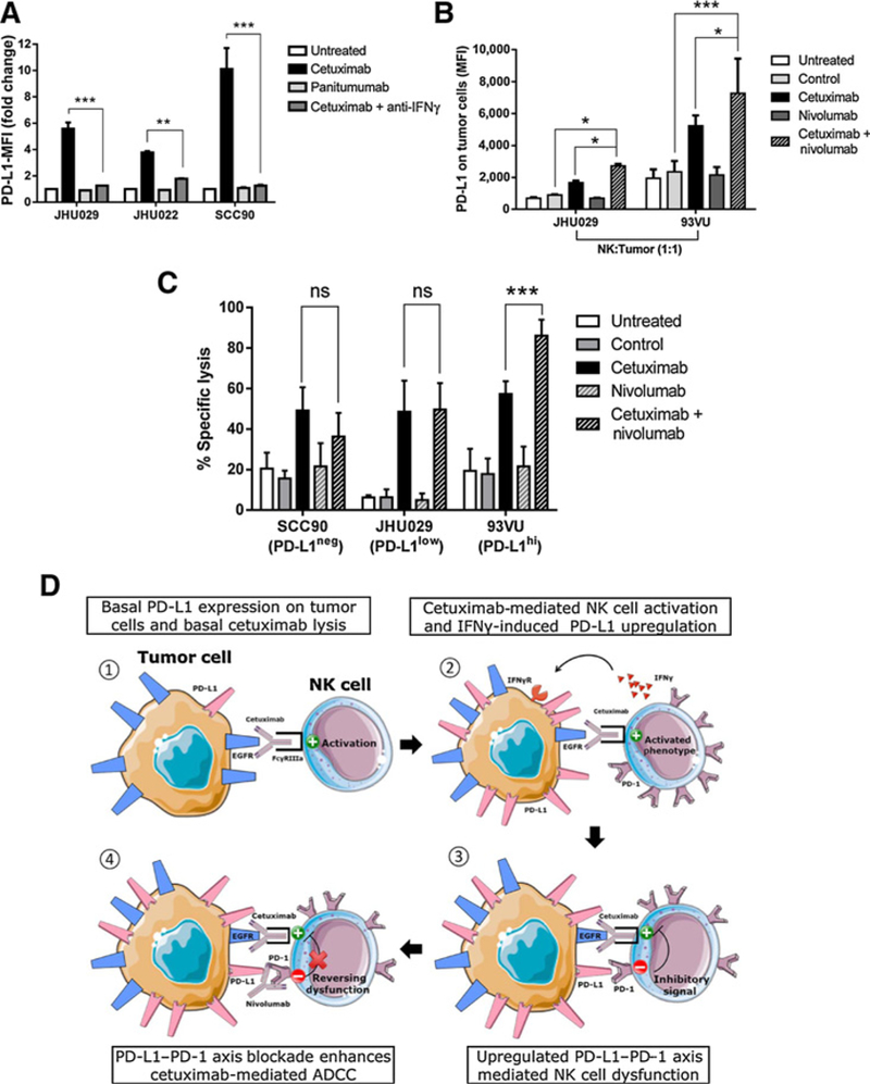
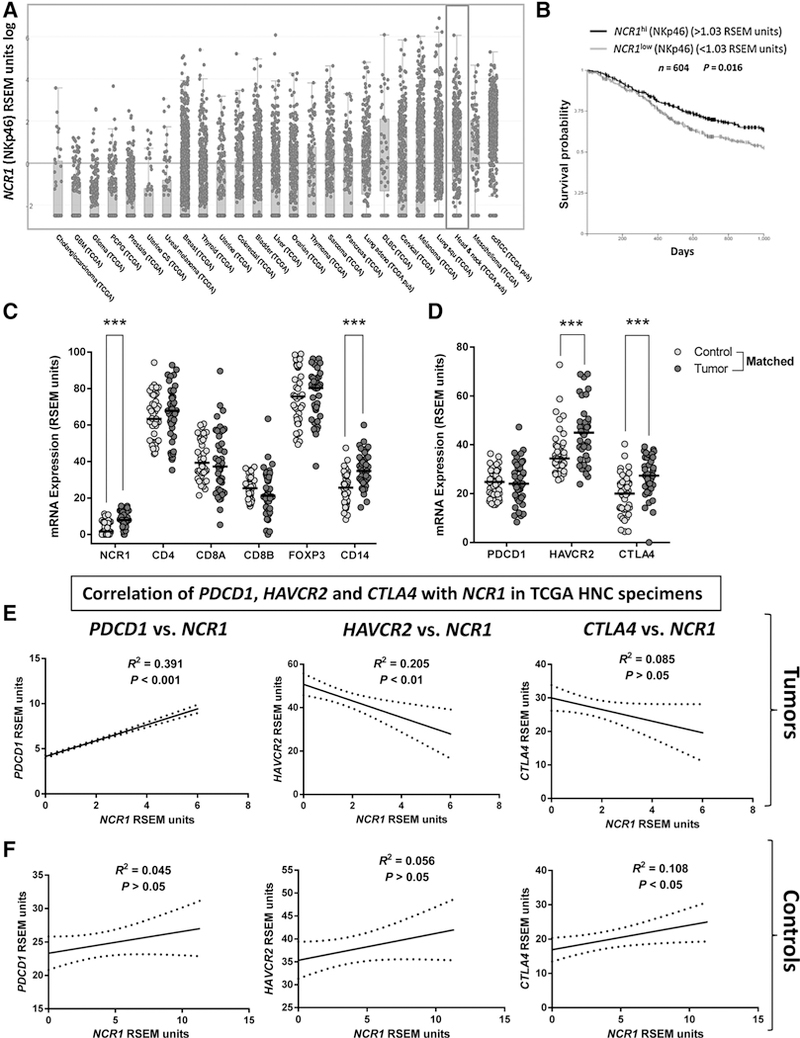
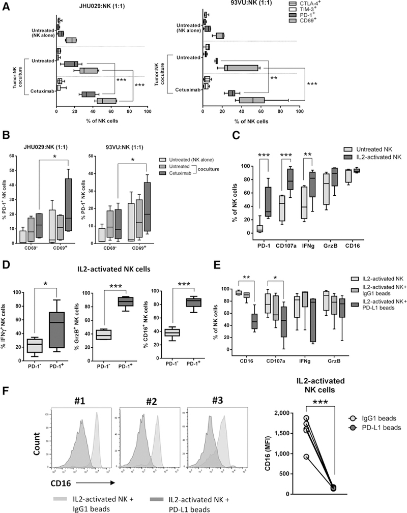
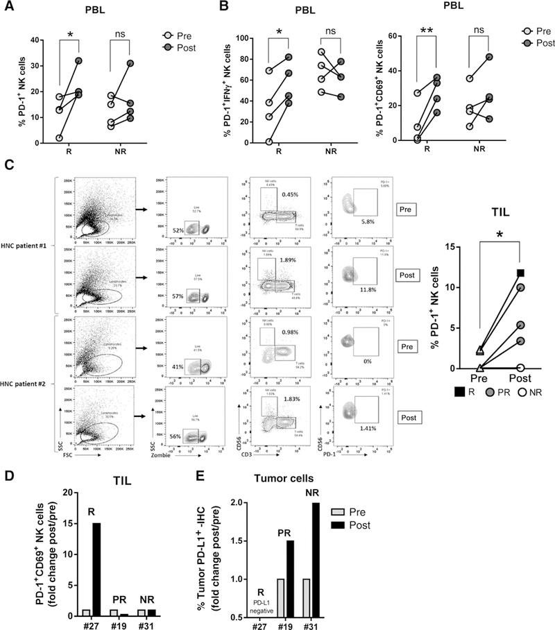
Inhibitory immune-checkpoint receptors (ICRs), including programmed death 1 (PD-1), have been characterized as exhaustion markers on T cells that infiltrate the tumor microenvironment (TME) of many cancer types, including head and neck cancer (HNC). However, expression and function of ICRs, including PD-1, on natural killer (NK) cells remains less defined. NK cells are innate immune effector cells that lyse epidermal growth factor receptor-overexpressing HNC cells via cetuximab-mediated antibody-dependent cytotoxicity. Cetuximab is clinically effective but only in 10% to 15% of patients. Therefore, it is necessary to investigate how immunomodulation with cetuximab or PD-1 blockade might enhance NK cell responses in the TME and improve monoclonal antibody therapeutic efficacy. We observed that expression of PD-1 on NK cells marks an activated phenotype, which was suppressed only after binding programmed death ligand-1 (PD-L1). HNC patients who exhibit higher circulating PD-1 NK cells associate with better clinical outcome, and these cells are enriched in the TME. Cetuximab-mediated NK cell activation increased PD-1 expression on NK cells which was confirmed in a prospective neoadjuvant cetuximab trial. In contrast, PD-L1 ligation of PD-1 NK cells diminished their activation status, whereas PD-1 blockade increased cetuximab-mediated NK cell activation and cytotoxicity, but only against HNC targets with high PD-L1 expression. Therefore, blocking the PD-1-PD-L1 axis may be a useful strategy to reverse immune evasion of HNC tumors with high PD-L1 expression during cetuximab therapy by reversing NK cell dysfunction.
抑制性免疫检查点受体(ICRs),包括程序性死亡受体 1(PD-1),已被鉴定为浸润多种癌症类型肿瘤微环境(TME)的 T 细胞的衰竭标志物,包括头颈部癌症(HNC)。然而,自然杀伤(NK)细胞上的 ICRs,包括 PD-1,的表达和功能仍不明确。NK 细胞是先天免疫效应细胞,通过西妥昔单抗介导的抗体依赖性细胞毒性裂解表皮生长因子受体过表达的 HNC 细胞。西妥昔单抗在临床上是有效的,但只有 10%到 15%的患者有效。因此,有必要研究西妥昔单抗或 PD-1 阻断如何通过免疫调节增强 TME 中的 NK 细胞反应并提高单克隆抗体治疗效果。我们观察到,NK 细胞上 PD-1 的表达标志着一种激活表型,只有在与程序性死亡配体-1(PD-L1)结合后才会受到抑制。表现出更高循环 PD-1 NK 细胞的 HNC 患者与更好的临床结果相关,并且这些细胞在 TME 中富集。西妥昔单抗介导的 NK 细胞激活增加了 NK 细胞上 PD-1 的表达,这在一项前瞻性新辅助西妥昔单抗试验中得到了证实。相比之下,PD-L1 结合 PD-1 NK 细胞会降低其激活状态,而 PD-1 阻断则会增加西妥昔单抗介导的 NK 细胞激活和细胞毒性,但仅针对 PD-L1 表达高的 HNC 靶标。因此,阻断 PD-1-PD-L1 轴可能是一种有用的策略,通过逆转 NK 细胞功能障碍来逆转高 PD-L1 表达的 HNC 肿瘤在西妥昔单抗治疗中的免疫逃逸。