Department of Clinical Radiology, Graduate School of Medical Sciences, Kyushu University.
Innovation Center for Medical Redox Navigation, Kyushu University.
Magn Reson Med Sci. 2019 Apr 10;18(2):142-149. doi: 10.2463/mrms.mp.2017-0157. Epub 2018 Oct 2.
To investigate the binding potential of newly developed Annexin V-conjugated ultrasmall superparamagnetic iron oxide (V-USPIO) for detection of drug-induced apoptosis in vitro and in vivo.
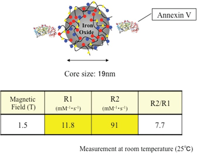
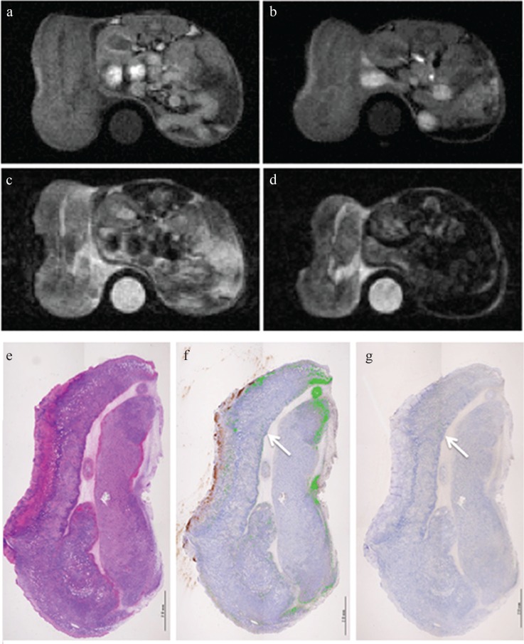
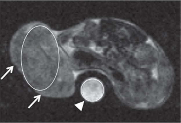
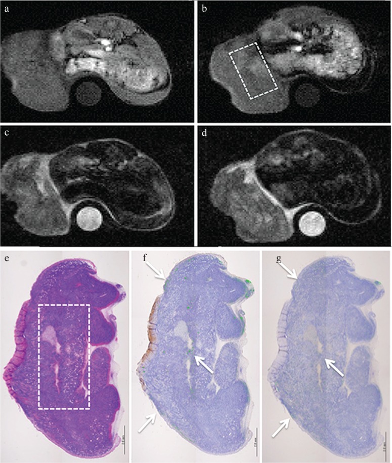
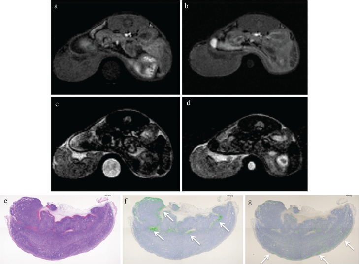
Apoptotic cells induced by camptothecin were incubated with or without Annexin V-USPIO at a concentration of 0.089 mmol Fe/L in vitro. T values of the two cell suspensions were measured by 0.47T nuclear magnetic resonance (NMR) spectrometer. Tumor-bearing mice were subjected to 1.5T MR scanner at 2 h after intraperitoneal injection of etoposide and cyclophosphamide. Following the pre-contrast T- and T-weighted imaging (0 h), the post-contrast scan was performed at 2, 4, 6 and 24 h after intravenous injection of Annexin V-USPIO (100 μmol Fe/kg). As a control, MRI was also obtained at 4 h after injection of USPIO without Annexin V. The ratio of tumor signal intensity (SI) on post-MRI for that on pre-MRI (Post/Pre-SI ratio) was calculated. After scanning, tumors were resected for pathological analysis to evaluate the distribution of iron and apoptotic cells.
The suspension of apoptotic cells incubated with Annexin V-USPIO showed shorter T value than that without it. On T-weighted imaging post/pre-SI ratio at 4 h after injection of Annexin V-USPIO showed 1.46, while after injection of USPIO without Annexin V was 1.17. The similar distribution of iron and apoptotic cells was observed in concordance with high signal intensity area on post-T-weighted imaging.
A newly developed Annexin V-USPIO could have the potential for detection of drug-induced apoptosis.
研究新开发的膜联蛋白 V 结合物超顺磁性氧化铁(V-USPIO)用于体外和体内检测药物诱导细胞凋亡的结合潜能。
用浓度为 0.089mmolFe/L 的膜联蛋白 V-USPIO 孵育体外培养的由喜树碱诱导的凋亡细胞或不与膜联蛋白 V-USPIO 孵育的凋亡细胞。用 0.47T 核磁共振(NMR)谱仪测量两种细胞悬液的 T 值。荷瘤小鼠在顺铂和环磷酰胺腹腔注射后 2h 行 1.5T 磁共振扫描仪检查。行对比前 T1 加权和 T2 加权成像(0h)后,静脉注射膜联蛋白 V-USPIO(100μmolFe/kg)后 2、4、6 和 24h 行对比后扫描。作为对照,在没有膜联蛋白 V 的 USPIO 注射后 4h 也进行 MRI 检查。计算 MRI 后肿瘤信号强度(SI)与 MRI 前肿瘤信号强度的比值(Post/Pre-SI 比值)。扫描后切除肿瘤,进行病理分析以评估铁和凋亡细胞的分布。
与没有膜联蛋白 V-USPIO 孵育的凋亡细胞悬液相比,孵育有膜联蛋白 V-USPIO 的凋亡细胞悬液的 T 值更短。在注射膜联蛋白 V-USPIO 后 4h,T1 加权成像后/前 SI 比值为 1.46,而注射无膜联蛋白 V-USPIO 后为 1.17。T1 加权成像后高信号强度区域的变化与铁和凋亡细胞的分布一致。
新开发的膜联蛋白 V-USPIO 具有检测药物诱导细胞凋亡的潜力。