Rissardo Jamir Pitton, Caprara Ana Letícia Fornari
Department of Neurology, Federal University of Santa Maria, Santa Maria, RS, Brazil.
Asian J Neurosurg. 2018 Jul-Sep;13(3):893-896. doi: 10.4103/ajns.AJNS_94_18.
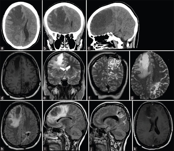
Tumefactive multiple sclerosis (MS) is characterized by the presence of a single MS-plaque in the brain. It mimics tumors due to large size, mass effect, and enhancement patterns. Refractory intracranial hypertension due to tumefactive MS requiring decompressive craniectomy (DC) was reported in five cases. However, none of these cases were documented new lesions during the follow-up. We report a case of a 28-year-old female admitted with acute right hemiparesis, headache, and nausea. A brain magnetic resonance imaging (MRI) revealed a left parietal lobe lesion. Within 4 days, she became comatose. Computed tomography (CT) scan revealed the left uncal herniation. DC and resection of the lesion were carried out. Histopathology revealed active demyelinating disease. After 11 years of the first attack, she went to the emergency department due to headache and left hemiparesis. Head CT scan revealed a hypodense area in the right frontal lobe. Three months later, the patient was asymptomatic, and new MRI did not show new lesions.
肿胀型多发性硬化症(MS)的特征是大脑中存在单个MS斑块。由于其体积大、占位效应和强化模式,它类似肿瘤。有5例报告了因肿胀型MS导致难治性颅内高压而需要进行减压颅骨切除术(DC)的情况。然而,这些病例在随访期间均未记录到新的病变。我们报告一例28岁女性,因急性右半身轻瘫、头痛和恶心入院。脑部磁共振成像(MRI)显示左顶叶有病变。4天内,她陷入昏迷。计算机断层扫描(CT)显示左侧钩回疝。进行了DC和病变切除。组织病理学显示为活动性脱髓鞘疾病。首次发作11年后,她因头痛和左半身轻瘫前往急诊科。头部CT扫描显示右额叶有一个低密度区。三个月后,患者无症状,新的MRI未显示新的病变。