Yan Bing, Fu Tong, Liu Yueming, Wei Wenbin, Dai Haojie, Fang Wei, Wang Feng
Beijing Key Laboratory of Nasal Diseases, Beijing Institute of Otolaryngology.
Department of Nuclear Medicine, Beijing Tongren Hospital, Capital Medical University, Beijing, China.
Medicine (Baltimore). 2018 Oct;97(40):e12441. doi: 10.1097/MD.0000000000012441.
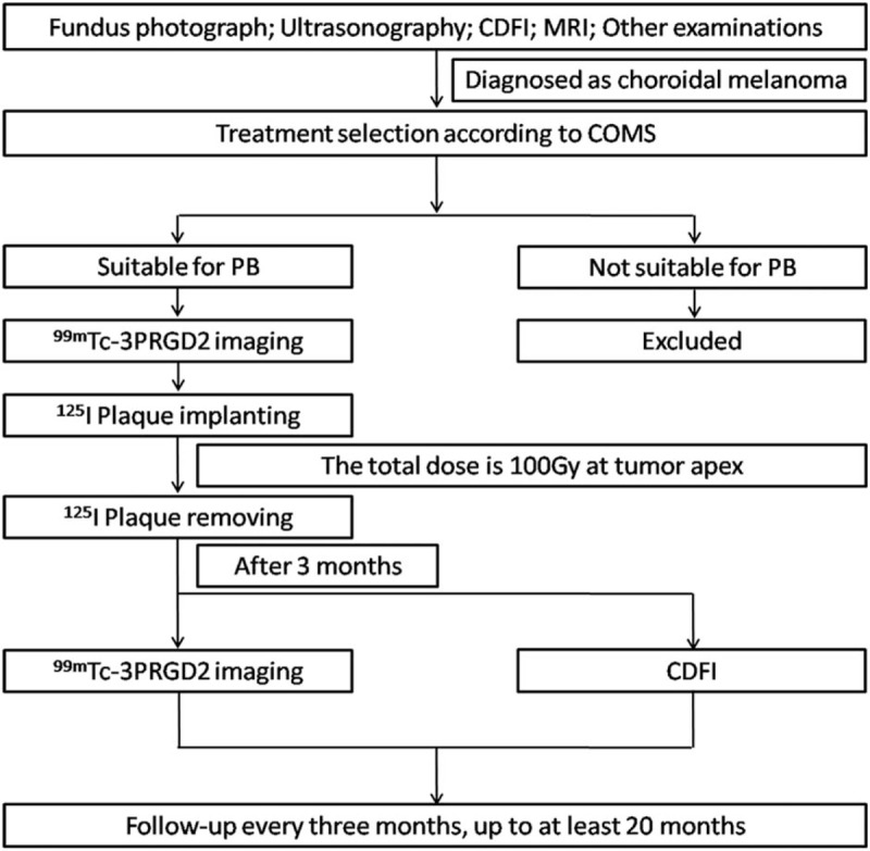
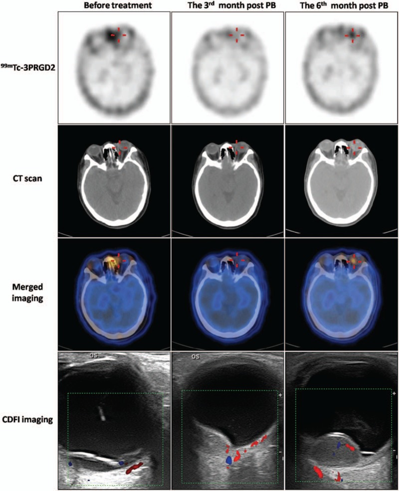
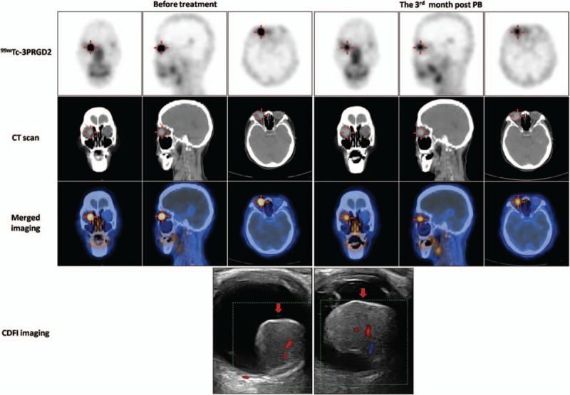
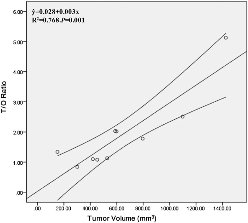
Recent successes in monitoring and diagnosing a variety of tumors using Tc-PEG4-E[PEG4-c(RGDfK)]2 (Tc-3PRGD2) single-photon emission computed tomography (SPECT) imaging encouraged us to expand the use of this tracer. This case-control study aimed to evaluate the feasibility of Tc-3PRGD2 imaging for detecting choroidal melanoma (CM) and for monitoring tumor response to plaque brachytherapy (PB). Ten consecutive patients with CM who underwent Tc-3PRGD2 imaging before and 3 months after PB were reviewed. The tumor-to-occipital bone (T/O) and mirrored contralateral normal tissue-to-occipital bone (N/O) ratios were calculated by region of interest analysis at baseline and 3 months post-PB. T/O values were compared between patients with CM with comorbid secondary retinal detachment (RD) and those without RD. The relationship between T/O value and tumor volume was also investigated. Tc-3PRGD2 SPECT/CT showed focal uptake in CM. The mean T/O ratio before PB was 1.90 ± 1.26 and the mean N/O ratio was 0.80 ± 0.21 (P = .02). The Tc-3PRGD2 concentrations in 5 patients with CM with RD were higher (T/O = 2.69 ± 1.39) than in those without secondary RD (T/O = 1.10 ± 0.18) (P = .008). T/O ratios at 3 months post-PB were significantly lower than that at baseline (1.23 ± 0.59, P = .03). There was a linear relationship between T/O and tumor volume (y-hat = 0.028 + 0.003x, R = 0.768, P = .001). The 95% confidence interval for the (T/O)/volume ratio was 0.002 to 0.005. Tc-3PRGD2 imaging is a feasible modality for the diagnosis of CM. Furthermore, follow-up for at least 20 months after PB indicated that coanalysis of Tc-3PRGD2 imaging and tumor volume may provide a promising prognostic predictor in patients with CM.
近期,使用锝标记的聚乙二醇-4-环[聚乙二醇-4-c(RGDfK)]2(Tc-PEG4-E[PEG4-c(RGDfK)]2,即Tc-3PRGD2)单光子发射计算机断层扫描(SPECT)成像技术在多种肿瘤的监测和诊断方面取得的成功,促使我们扩大这种示踪剂的应用。本病例对照研究旨在评估Tc-3PRGD2成像用于检测脉络膜黑色素瘤(CM)以及监测肿瘤对敷贴近距离放射治疗(PB)反应的可行性。回顾了10例连续的CM患者,他们在PB治疗前和治疗后3个月均接受了Tc-3PRGD2成像检查。通过感兴趣区分析计算基线期和PB治疗后3个月时肿瘤与枕骨的比值(T/O)以及对侧镜像正常组织与枕骨的比值(N/O)。比较了合并继发性视网膜脱离(RD)的CM患者和未合并RD的CM患者之间的T/O值。还研究了T/O值与肿瘤体积之间的关系。Tc-3PRGD2 SPECT/CT显示CM有局灶性摄取。PB治疗前的平均T/O比值为1.90±1.26,平均N/O比值为0.80±0.21(P = 0.02)。5例合并RD的CM患者的Tc-3PRGD2浓度(T/O = 2.69±1.39)高于未合并继发性RD的患者(T/O = 1.10±0.18)(P = 0.008)。PB治疗后3个月时的T/O比值显著低于基线期(1.23±0.59,P = 0.03)。T/O与肿瘤体积之间存在线性关系(y-hat = 0.028 + 0.003x,R = 0.768,P = 0.001)。(T/O)/体积比值的95%置信区间为0.002至0.005。Tc-3PRGD2成像对于CM的诊断是一种可行的方法。此外,PB治疗后至少20个月的随访表明,Tc-3PRGD2成像与肿瘤体积的联合分析可能为CM患者提供一个有前景的预后预测指标。