da Silva Nivaldo Adolfo, Vassallo José, Sarian Luis Otávio, Cognard Christophe, Sevely Annick
Department of Radiology, Faculty of Medical Sciences, State University of Campinas, Campinas (UNICAMP), Campinas-SP, Brazil.
Department of Diagnostic and Therapeutic Neuroradiology, Centre Hospitalier Universitaire (CHU) - Hôpital Pierre Paul Riquet, University of Toulouse, Toulouse, France.
Medicine (Baltimore). 2018 Oct;97(40):e12602. doi: 10.1097/MD.0000000000012602.
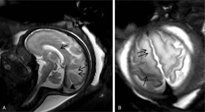
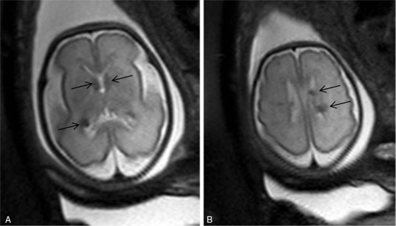
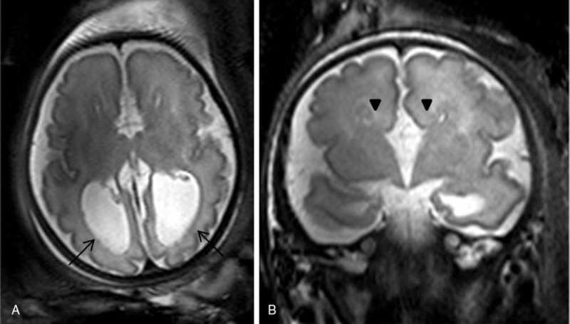
To report our preliminary experience with cerebral fetal magnetic resonance imaging (MRI) with a 3 Tesla (3T) scanner. We assessed feasibility, time of acquisition, and possibility to establish a diagnosis.Fifty-nine pregnant women had fetal MRI performed during the third trimester of pregnancy due to clinical or sonography concern of a central nervous system anomaly. No fetal or maternal sedation was used. The MRI protocol consisted of T2 turbo-spin-echo images in 3 planes of space. No T1-weighted images were performed. All images were analyzed by 2 pediatric neuroradiologists, who evaluated spatial resolution, artifacts, time of acquisition, and possibility to establish a diagnosis suspected by sonography.Examinations were performed safely for all patients. The images required longer time of acquisition (approximately 75 seconds for each plane in the space). The specific absorption rate was not exceeded in any fetus. Cerebral fetal MRI was normal in 22 cases. The spectrum of diagnostics included isolated ventriculomegaly, posterior fossa malformation, corpus callosum malformation, gyration anomalies, craniosynostosis, tuberous sclerosis, microcephaly, external hydrocephaly, midline arachnoid cyst, cerebral lesions, and persistent hyperplastic primitive vitreous.In our series, 3 T MRI of fetal brain was feasible and able to establish a diagnosis but required longer time of acquisition.
报告我们使用3特斯拉(3T)扫描仪进行胎儿脑磁共振成像(MRI)的初步经验。我们评估了可行性、采集时间以及做出诊断的可能性。59名孕妇在妊娠晚期因临床或超声检查怀疑存在中枢神经系统异常而接受了胎儿MRI检查。未使用胎儿或母体镇静剂。MRI方案包括在三个空间平面上的T2快速自旋回波图像。未进行T1加权图像检查。所有图像由两名儿科神经放射科医生进行分析,他们评估了空间分辨率、伪影、采集时间以及做出超声检查怀疑诊断的可能性。所有患者的检查均安全进行。图像需要更长的采集时间(空间中每个平面约75秒)。任何胎儿的比吸收率均未超过。22例胎儿脑MRI结果正常。诊断范围包括孤立性脑室扩大、后颅窝畸形、胼胝体畸形、脑回异常、颅骨缝早闭、结节性硬化症、小头畸形、外部脑积水、中线蛛网膜囊肿、脑病变以及持续性增生性原始玻璃体。在我们的系列研究中,胎儿脑3T MRI可行且能够做出诊断,但需要更长的采集时间。