• 文献检索
  • 文档翻译
  • 深度研究
  • 学术资讯
  • Suppr Zotero 插件Zotero 插件
  • 邀请有礼
  • 套餐&价格
  • 历史记录
应用&插件
Suppr Zotero 插件Zotero 插件浏览器插件Mac 客户端Windows 客户端微信小程序
定价
高级版会员购买积分包购买API积分包
服务
文献检索文档翻译深度研究API 文档MCP 服务
关于我们
关于 Suppr公司介绍联系我们用户协议隐私条款
关注我们

Suppr 超能文献

核心技术专利:CN118964589B侵权必究
粤ICP备2023148730 号-1Suppr @ 2026

文献检索

告别复杂PubMed语法,用中文像聊天一样搜索,搜遍4000万医学文献。AI智能推荐,让科研检索更轻松。

立即免费搜索

文件翻译

保留排版,准确专业,支持PDF/Word/PPT等文件格式,支持 12+语言互译。

免费翻译文档

深度研究

AI帮你快速写综述,25分钟生成高质量综述,智能提取关键信息,辅助科研写作。

立即免费体验

正颌手术后软组织的三维评估。

Three dimensional evaluation of soft tissue after orthognathic surgery.

机构信息

Department of Oral and Maxillofacial Surgery, School of Dentistry, Kyung Hee University, 26, Kyungheedae-ro, Dongdaemun-gu, 02447, Seoul, Republic of Korea.

出版信息

Head Face Med. 2018 Oct 5;14(1):21. doi: 10.1186/s13005-018-0179-z.

DOI:10.1186/s13005-018-0179-z
PMID:30290762
原文链接:https://pmc.ncbi.nlm.nih.gov/articles/PMC6173856/
Abstract

BACKGROUND

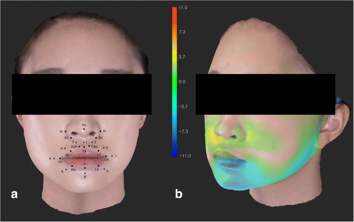
To evaluate the nasolabial soft tissue change three-dimensionally after orthognathic surgery, using a structured light scanner.

METHODS

Thirty-two malocclusion patients, who underwent orthognathic surgery, were evaluated. CBCT and 3D facial scans were obtained before surgery and 3 months after surgery. The 3D changes in the 26 landmarks, and the relative ratio of the soft tissue movement to the bony movement, were evaluated.

RESULTS

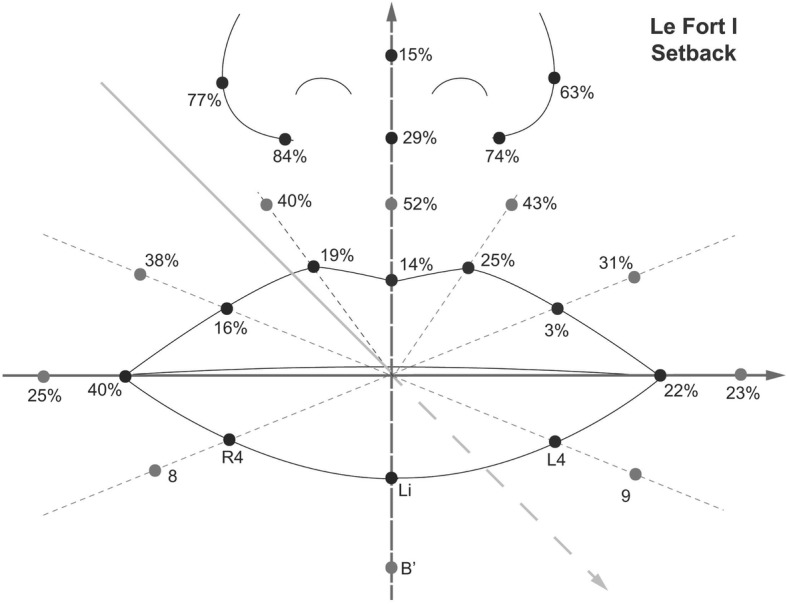
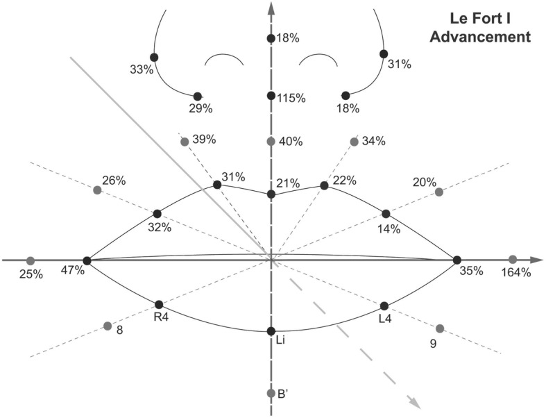
In the Le Fort I advancement patients, the nasal tip moved 17% forward, compared to the maxillary bony movement, but the nasal prominence decreased 15%. The alar width increased 4 mm after the advancement, and the width decreased 4.7 mm after Le Fort I setback. The relative ratio of the soft tissue movement to the bony movement after bilateral sagittal split osteotomy was about 66% at the Li point in the anteroposterior direction, and it was 21% in the Le Fort I advancement and 14% in Le Fort I setback at the Ls point.

CONCLUSION

Alar cinch suturing may not be sufficient to overcome the effect of the maxilla advancement compressing the nasal complex. Alar width widening was prevented in Le Fort I setback. However, it is uncertain that the alar cinch suturing was solely responsible. The soft tissue around the mandible tends to accompany the bony movement more than the maxillary area. In addition, structured light scanning system proved to be a useful tool to evaluate the nasolabial soft tissue.

摘要

背景

使用结构光扫描仪评估正颌手术后的鼻唇软组织的三维变化。

方法

对 32 例接受正颌手术的错畸形患者进行评估。在术前和术后 3 个月分别获得 CBCT 和 3D 面部扫描。评估了 26 个标志点的 3D 变化以及软组织运动与骨运动的相对比率。

结果

在 Le Fort I 前徙患者中,与上颌骨的骨运动相比,鼻尖向前移动了 17%,但鼻突却减少了 15%。前徙后鼻翼宽度增加了 4mm,Le Fort I 后退后宽度减少了 4.7mm。双侧矢状劈开截骨术后,Li 点前后向的软组织运动与骨运动的相对比率约为 66%,Le Fort I 前徙和 Le Fort I 后退时 Ls 点的比率分别为 21%和 14%。

结论

鼻翼缩窄缝合可能不足以克服上颌前徙对鼻复合体的压缩作用。Le Fort I 后退术可防止鼻翼增宽。然而,鼻翼缩窄缝合是否是唯一的原因尚不确定。下颌周围的软组织往往比上颌区域更伴随骨运动。此外,结构光扫描系统被证明是评估鼻唇软组织的有用工具。

https://cdn.ncbi.nlm.nih.gov/pmc/blobs/c344/6173856/ed767861476c/13005_2018_179_Fig4_HTML.jpg
https://cdn.ncbi.nlm.nih.gov/pmc/blobs/c344/6173856/e266366698c2/13005_2018_179_Fig1_HTML.jpg
https://cdn.ncbi.nlm.nih.gov/pmc/blobs/c344/6173856/bc3b49c1c653/13005_2018_179_Fig2_HTML.jpg
https://cdn.ncbi.nlm.nih.gov/pmc/blobs/c344/6173856/222f80abb61a/13005_2018_179_Fig3_HTML.jpg
https://cdn.ncbi.nlm.nih.gov/pmc/blobs/c344/6173856/ed767861476c/13005_2018_179_Fig4_HTML.jpg

相似文献

1
Three dimensional evaluation of soft tissue after orthognathic surgery.正颌手术后软组织的三维评估。
Head Face Med. 2018 Oct 5;14(1):21. doi: 10.1186/s13005-018-0179-z.
2
Effect of setback Le Fort I osteotomy on midfacial soft-tissue changes as evaluated by cone-beam computed tomography superimposition for cases of skeletal Class III malocclusion.通过锥形束计算机断层扫描叠加评估骨骼 III 类错畸形病例中后退性 Le Fort I 截骨术对上颌骨软组织变化的影响。
Int J Oral Maxillofac Surg. 2013 Jun;42(6):790-5. doi: 10.1016/j.ijom.2012.11.012. Epub 2013 Jan 5.
3
Three-dimensional changes in nose and upper lip volume after orthognathic surgery.正颌外科手术后鼻和上唇体积的三维变化
Int J Oral Maxillofac Surg. 2015 Jan;44(1):83-9. doi: 10.1016/j.ijom.2014.08.001. Epub 2014 Sep 11.
4
The evaluation of the nasal morphologic changes after bimaxillary surgery in skeletal class III maloccusion by using the superimposition of cone-beam computed tomography (CBCT) volumes.应用锥形束 CT 容积叠加技术评价骨性 III 类错[牙合]患者双颌手术后的鼻形态变化。
J Craniomaxillofac Surg. 2012 Jun;40(4):e87-92. doi: 10.1016/j.jcms.2011.05.008. Epub 2011 Jul 2.
5
Three-dimensional evaluation of the alar cinch suture after Le Fort I osteotomy.Le Fort I型截骨术后鼻翼缩紧缝合的三维评估
Int J Oral Maxillofac Surg. 2016 Oct;45(10):1309-14. doi: 10.1016/j.ijom.2016.05.013. Epub 2016 Jun 3.
6
Three-Dimensional Analysis of Midfacial Soft Tissue Changes After Maxillary Posterior Impaction and Intraoral Vertical Ramus Osteotomy for Mandibular Setback in Class III Patients.Ⅲ类错颌患者上颌后份垂直牵引及下颌升支矢状劈开截骨后退术后面中份软组织变化的三维分析
J Craniofac Surg. 2017 Oct;28(7):1789-1796. doi: 10.1097/SCS.0000000000003754.
7
Midfacial soft-tissue changes after advancement of maxilla with Le Fort I osteotomy and mandibular setback surgery: comparison of conventional and high Le Fort I osteotomies by superimposition of cone-beam computed tomography volumes.采用Le Fort I型截骨术推进上颌骨及下颌后退手术后的面中部软组织变化:通过锥形束计算机断层扫描容积叠加对比传统Le Fort I型截骨术和高位Le Fort I型截骨术
J Oral Maxillofac Surg. 2011 Jun;69(6):e225-33. doi: 10.1016/j.joms.2010.12.035. Epub 2011 Apr 15.
8
[Effect of alar base cinch suture based on anatomic landmarks on the morphology of nasolabial region in patients after orthognathic surgery].[基于解剖标志的鼻翼基底收紧缝合对正颌外科术后患者鼻唇区域形态的影响]
Beijing Da Xue Xue Bao Yi Xue Ban. 2023 Aug 18;55(4):736-742. doi: 10.19723/j.issn.1671-167X.2023.04.027.
9
Nasal Soft Tissue Change Following Bimaxillary Orthognathic Surgery.双颌正颌外科手术后鼻软组织的变化
J Craniofac Surg. 2017 Oct;28(7):e605-e608. doi: 10.1097/SCS.0000000000003736.
10
Three-dimensional changes in the location of soft tissue landmarks following bimaxillary orthognathic surgery.双颌正颌手术后软组织标志点位置的三维变化。
J Orofac Orthop. 2021 Jul;82(4):257-265. doi: 10.1007/s00056-021-00279-1. Epub 2021 Mar 25.

引用本文的文献

1
Comparison of soft tissue effects of different orthognathic surgical approaches in class III patients using photographic analysis.使用摄影分析比较Ⅲ类患者不同正颌外科手术方法对软组织的影响。
Sci Rep. 2025 Apr 30;15(1):15204. doi: 10.1038/s41598-025-00020-7.
2
Optical Three-Dimensional Imaging for Objective Evaluation of the Donor Site after Anterolateral Thigh Flap Surgery.光学三维成像用于客观评估股前外侧皮瓣手术后供区情况
J Clin Med. 2024 Mar 21;13(6):1805. doi: 10.3390/jcm13061805.
3
Effects of Maxillary Movements on Lips Following Orthognathic Surgery: A Retrospective Non-Randomized Clinical Trial.

本文引用的文献

1
Assessment of Alar Flare and Efficacy of Alar Cinch Suture in the Management of Alar Flare Following Le Fort 1 Superior Repositioning: A Comparative Study.勒福Ⅰ型上颌骨上移术后鼻翼外扩的评估及鼻翼缩窄缝合术治疗鼻翼外扩的疗效:一项对比研究
J Maxillofac Oral Surg. 2016 Dec;15(4):528-534. doi: 10.1007/s12663-015-0866-2. Epub 2015 Dec 14.
2
A systematic review of soft-to-hard tissue ratios in orthognathic surgery. Part III: Double jaw surgery procedures.正颌外科软硬组织比例的系统评价。第三部分:双颌手术程序。
J Craniomaxillofac Surg. 2016 Oct;44(10):1599-1606. doi: 10.1016/j.jcms.2016.08.016. Epub 2016 Aug 21.
3
正颌外科手术后上颌移动对唇部的影响:一项回顾性非随机临床试验
Int Arch Otorhinolaryngol. 2024 Feb 5;28(1):e148-e156. doi: 10.1055/s-0043-1773759. eCollection 2024 Jan.
4
Clockwise versus counterclockwise rotation in bimaxillary surgery: 3D analysis of facial soft tissue outcomes.双颌手术中顺时针与逆时针旋转:面部软组织结果的 3D 分析。
Oral Maxillofac Surg. 2024 Jun;28(2):693-703. doi: 10.1007/s10006-023-01196-w. Epub 2023 Nov 20.
5
Application of three-dimensional reconstruction technology in dentistry: a narrative review.三维重建技术在牙科中的应用:叙述性综述。
BMC Oral Health. 2023 Sep 4;23(1):630. doi: 10.1186/s12903-023-03142-4.
6
The Use of 3D Technology in the Management of Residual Asymmetry following Orthognathic Surgery: A Case Report.3D技术在正颌外科术后残余不对称管理中的应用:一例报告
Healthcare (Basel). 2023 Jul 31;11(15):2172. doi: 10.3390/healthcare11152172.
7
Analysis of select esthetic nasal parameters in virtually planned orthognathic patients.虚拟计划正颌患者的部分美学鼻部参数分析。
Eur Arch Otorhinolaryngol. 2023 Aug;280(8):3885-3890. doi: 10.1007/s00405-023-08031-8. Epub 2023 May 25.
8
Orthognathic Surgery as Class III Skeletal Treatment in a 31-Year-Old Female with Mandible Prognathism: A Case Report.正颌外科手术治疗一名31岁下颌前突女性Ⅲ类骨骼畸形:病例报告
Eur J Dent. 2023 Jul;17(3):935-942. doi: 10.1055/s-0043-1761453. Epub 2023 Mar 28.
9
3-dimensional analysis of nasal soft tissue alterations following maxillary Lefort I advancement with and without impaction using 3D photogrammetry scanner.上颌骨 Le Fort I 推进术伴或不伴压入的三维摄影扫描仪测量的鼻软组织变化的三维分析。
Oral Maxillofac Surg. 2024 Mar;28(1):111-123. doi: 10.1007/s10006-022-01121-7. Epub 2022 Nov 2.
10
Simultaneous PSI-Based Orthognathic and PEEK Bone Augmentation Surgery Leads to Improved Symmetric Facial Appearance in Craniofacial Malformations.基于PSI的正颌手术与PEEK骨增量手术同期进行可改善颅面畸形患者的面部对称性外观。
J Pers Med. 2022 Oct 5;12(10):1653. doi: 10.3390/jpm12101653.
Three-dimensional evaluation of the alar cinch suture after Le Fort I osteotomy.
Le Fort I型截骨术后鼻翼缩紧缝合的三维评估
Int J Oral Maxillofac Surg. 2016 Oct;45(10):1309-14. doi: 10.1016/j.ijom.2016.05.013. Epub 2016 Jun 3.
4
Accuracy and precision of integumental linear dimensions in a three-dimensional facial imaging system.三维面部成像系统中皮肤线性尺寸的准确性和精确性。
Korean J Orthod. 2015 May;45(3):105-12. doi: 10.4041/kjod.2015.45.3.105. Epub 2015 May 15.
5
Assessment of three-dimensional nasolabial response to Le Fort I advancement.评估 Le Fort I 提升术对鼻唇三维反应的影响。
J Plast Reconstr Aesthet Surg. 2014 Jun;67(6):756-63. doi: 10.1016/j.bjps.2014.03.023. Epub 2014 Apr 4.
6
Measurement of three-dimensional perioral soft tissue changes in dentoalveolar protrusion patients after orthodontic treatment using a structured light scanner.使用结构光扫描仪测量牙列前突患者正畸治疗后口周三维软组织变化。
Angle Orthod. 2014 Sep;84(5):795-802. doi: 10.2319/112913-877.1. Epub 2014 Mar 10.
7
Nasolabial soft tissue changes after Le Fort I advancement.Le Fort I型前徙术后鼻唇软组织变化
J Oral Maxillofac Surg. 2012 Apr;70(4):e270-7. doi: 10.1016/j.joms.2011.11.022. Epub 2012 Jan 28.
8
The influence of a Le Fort I impaction and advancement osteotomy on smile using a modified alar cinch suture and V-Y closure: a prospective study.Le Fort I 骨折伴推进截骨术对鼻唇沟改良固定缝线和 V-Y 缝合术微笑影响的前瞻性研究。
Int J Oral Maxillofac Surg. 2012 May;41(5):547-52. doi: 10.1016/j.ijom.2011.08.011. Epub 2011 Dec 29.
9
Nasal cavity volume changes after rapid maxillary expansion in adolescents evaluated with 3-dimensional simulation and modeling programs.青少年上颌快速扩弓后鼻腔容积的变化的 3 维模拟和建模程序评估。
Am J Orthod Dentofacial Orthop. 2011 Nov;140(5):633-40. doi: 10.1016/j.ajodo.2010.12.020.
10
Midfacial soft-tissue changes after advancement of maxilla with Le Fort I osteotomy and mandibular setback surgery: comparison of conventional and high Le Fort I osteotomies by superimposition of cone-beam computed tomography volumes.采用Le Fort I型截骨术推进上颌骨及下颌后退手术后的面中部软组织变化:通过锥形束计算机断层扫描容积叠加对比传统Le Fort I型截骨术和高位Le Fort I型截骨术
J Oral Maxillofac Surg. 2011 Jun;69(6):e225-33. doi: 10.1016/j.joms.2010.12.035. Epub 2011 Apr 15.