INSERM (French National Institute of Health and Medical Research), UMR 1260, Regenerative Nanomedicine (RNM), Fédération de Médecine Translationnelle de Strasbourg (FMTS), 11 rue Humann, Strasbourg, 67000, France.
Université de Strasbourg (UDS), Faculté de Chirurgie-dentaire, 8 rue Sainte-Elisabeth, Strasbourg, 67000, France.
Sci Rep. 2018 Oct 8;8(1):14914. doi: 10.1038/s41598-018-33267-4.
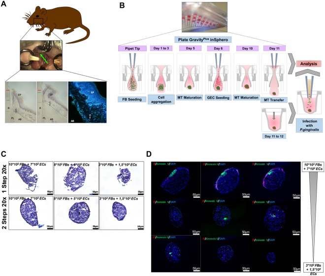
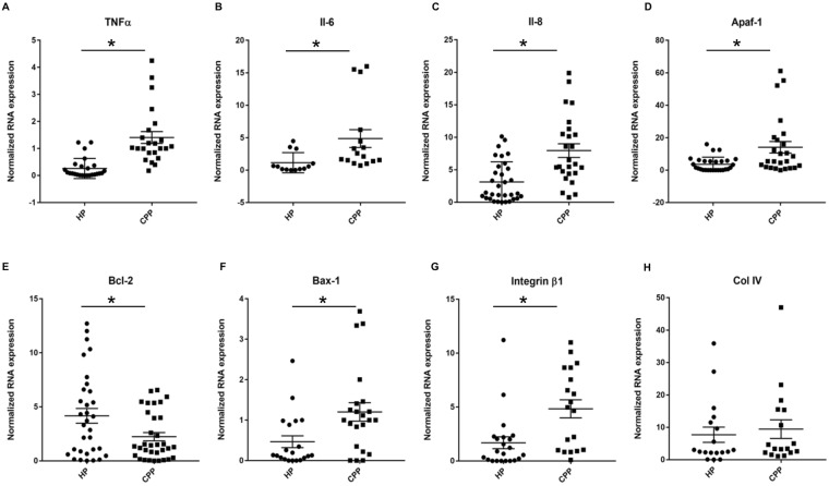
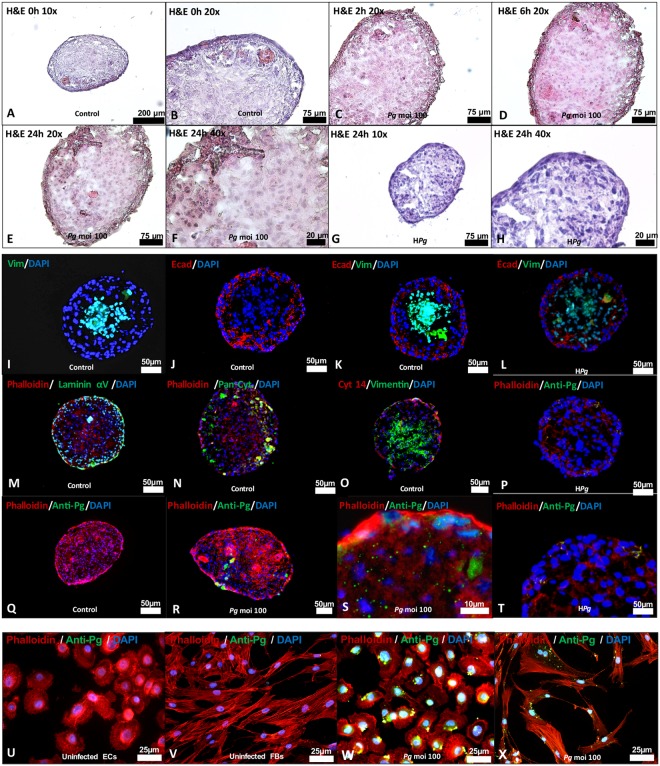
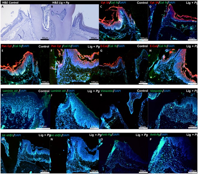
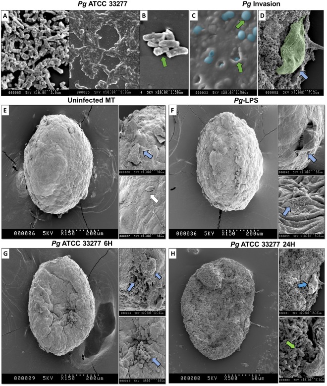
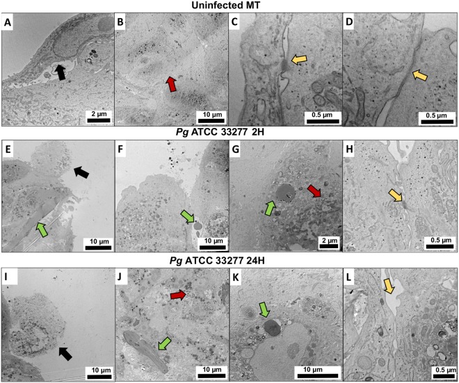
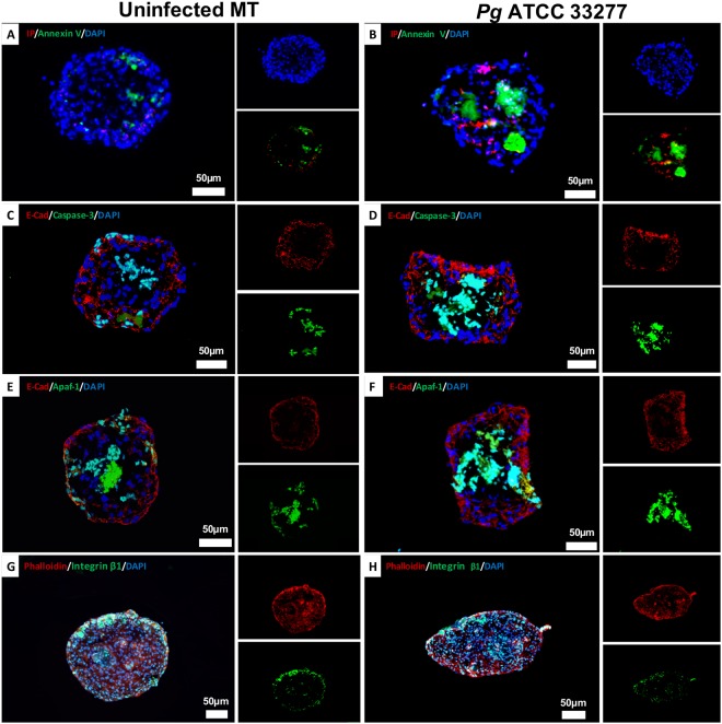
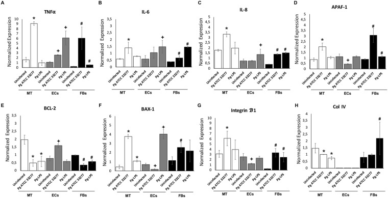
Porphyromonas gingivalis-induced inflammatory effects are mostly investigated in monolayer cultured cells. The aim of this study was to develop a 3D spheroid model of gingiva to take into account epithelio-fibroblastic interactions. Human gingival epithelial cells (ECs) and human oral fibroblasts (FBs) were cultured by hanging drop method to generate 3D microtissue (MT) whose structure was analyzed on histological sections and the cell-to-cell interactions were observed by scanning and transmission electron microscopy (SEM and TEM). MTs were infected by P. gingivalis and the impact on cell death (Apaf-1, caspase-3), inflammatory markers (TNF-α, IL-6, IL-8) and extracellular matrix components (Col-IV, E-cadherin, integrin β1) was evaluated by immunohistochemistry and RT-qPCR. Results were compared to those observed in situ in experimental periodontitis and in human gingival biopsies. MTs exhibited a well-defined spatial organization where ECs were organized in an external cellular multilayer, while, FBs constituted the core. The infection of MT demonstrated the ability of P. gingivalis to bypass the epithelial barrier in order to reach the fibroblastic core and induce disorganization of the spheroid structure. An increased cell death was observed in fibroblastic core. The development of such 3D model may be useful to define the role of EC-FB interactions on periodontal host-immune response and to assess the efficacy of new therapeutics.
牙龈卟啉单胞菌诱导的炎症反应主要在单层培养细胞中进行研究。本研究旨在建立一种考虑上皮-成纤维细胞相互作用的牙龈 3D 球体模型。通过悬滴法培养人牙龈上皮细胞(ECs)和人口腔成纤维细胞(FBs),生成 3D 微组织(MT),对其结构进行组织学切片分析,并通过扫描和透射电子显微镜(SEM 和 TEM)观察细胞间相互作用。用牙龈卟啉单胞菌感染 MT,通过免疫组织化学和 RT-qPCR 评估细胞死亡(凋亡相关因子 1(Apaf-1)、半胱天冬酶-3)、炎症标志物(肿瘤坏死因子-α(TNF-α)、白细胞介素-6(IL-6)、白细胞介素-8(IL-8))和细胞外基质成分(IV 型胶原(Col-IV)、E-钙黏蛋白、整合素β1)的变化。并将结果与实验性牙周炎和人牙龈活检中的观察结果进行比较。MT 显示出明确的空间组织,EC 排列在外层细胞的多层中,而 FB 构成核心。MT 的感染表明牙龈卟啉单胞菌能够绕过上皮屏障,到达成纤维细胞核心,并诱导球体结构的紊乱。在成纤维细胞核心观察到细胞死亡增加。这种 3D 模型的发展可能有助于确定 EC-FB 相互作用在牙周宿主免疫反应中的作用,并评估新疗法的疗效。