Onay Emel-Olga, Yamanel Kivanc, Korkmaz-Ceyhan Yonca, Gulsahi Kamran
Professor, Department of Endodontics, School of Dentistry, Baskent University, Bahcelievler-Ankara, Turkey.
Associate Professor, Department of Restorative Dentistry, School of Dentistry, Baskent University, Bahcelievler-Ankara, Turkey.
J Clin Exp Dent. 2018 Aug 1;10(8):e781-e788. doi: 10.4317/jced.54843. eCollection 2018 Aug.
Dental surface conditioning by Er:YAG laser is currently being investigated, as not all of the mechanisms and effects of this technique have been clearly studied. Thus, the aim of the present study was to assess the cervical microleakage of Class II resin composite restorations in endodontically treated teeth following either the respective conventional conditioning or additional Er:YAG laser conditioning, in association with varied adhesives.
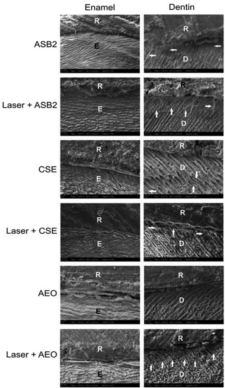
Standardized mesial-occlusal-distal cavities (two gingival walls positioned in dentin and enamel, respectively) were created in 60 extracted human premolar teeth. Following the completion of the endodontic therapy, the teeth were grouped into six categories based on conditioning modality and adhesive strategy as follows: group 1-37% phosphoric acid/Adper Single Bond 2 (ASB2); group 2-Er:YAG laser/37% phosphoric acid/ASB2; group 3-Clearfil SE Bond (CSE); group 4-Er:YAG laser/CSE; group 5-Adper Easy One (AEO); and group 6-Er:YAG laser/AEO. Specimens were submitted to thermocycling and dye penetration, followed by longitudinal sectioning. The dye penetration was evaluated using a stereomicroscope. One specimen from each group was assessed under a scanning electron microscope for adhesive interface analysis.
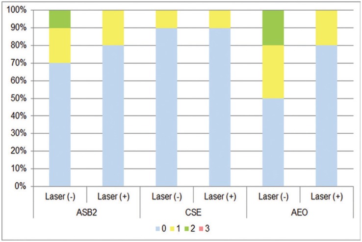
No significant differences were found between the conditioning modalities, nor between the adhesive systems at both margins. Groups 1 and 2 showed a lower degree of microleakage in the enamel vs. dentin ( = 0.002). Group 2 showed a significantly lower incidence of microleakage in enamel vs. dentin ( = 0.005).
CSE and AEO were comparable with that of ASB2 regarding sealing ability. Additional Er:YAG laser conditioning may be beneficial before ASB2 application in enamel. Endodontically treated teeth, etch-and-rinse adhesive, Er:YAG laser, gingival level, sealing ability, self-etch adhesive.
由于铒钇铝石榴石(Er:YAG)激光对牙面的预处理技术的所有机制和效果尚未得到明确研究,目前正在对其进行调查。因此,本研究的目的是评估在根管治疗后的牙齿中,采用相应的传统预处理或额外的Er:YAG激光预处理,并结合不同的粘结剂,Ⅱ类树脂复合材料修复体的颈部微渗漏情况。
在60颗拔除的人类前磨牙上制备标准化的近中-咬合-远中洞型(两个龈壁分别位于牙本质和釉质中)。根管治疗完成后,根据预处理方式和粘结策略将牙齿分为六组,如下:第1组-37%磷酸/Adper单键2(ASB2);第2组-Er:YAG激光/37%磷酸/ASB2;第3组-Clearfil SE粘结剂(CSE);第4组-Er:YAG激光/CSE;第5组-Adper简易一步法粘结剂(AEO);第6组-Er:YAG激光/AEO。将标本进行热循环和染料渗透试验,然后进行纵向切片。使用体视显微镜评估染料渗透情况。从每组中选取一个标本,在扫描电子显微镜下进行粘结界面分析。
在两种边缘情况下,预处理方式之间以及粘结系统之间均未发现显著差异。第1组和第2组在釉质中的微渗漏程度低于牙本质(P = 0.002)。第2组在釉质中的微渗漏发生率显著低于牙本质(P = 0.005)。
CSE和AEO在密封能力方面与ASB2相当。在应用ASB2之前,额外的Er:YAG激光预处理可能对釉质有益。根管治疗后的牙齿、酸蚀冲洗粘结剂、Er:YAG激光、龈缘水平、密封能力、自酸蚀粘结剂。