Yang Hsin-Yu, Yang Chang-Sue
Department of Ophthalmology, Taipei Veterans General Hospital, No. 201 Shipai Road, Sec. 2, Taipei, 11217, Taiwan.
Department of Ophthalmology, Shin-Kong Wu Ho-Su Memorial Hospital, No. 95, Wen Chang Road, Taipei, Taiwan.
BMC Ophthalmol. 2018 Oct 11;18(1):265. doi: 10.1186/s12886-018-0932-x.
To demonstrate a full thickness macular hole (MH) development after vitrectomy (VT) for rhegmatogenous retinal detachment (RRD) and to investigate the possible disease mechanism with optical coherence tomography (OCT).
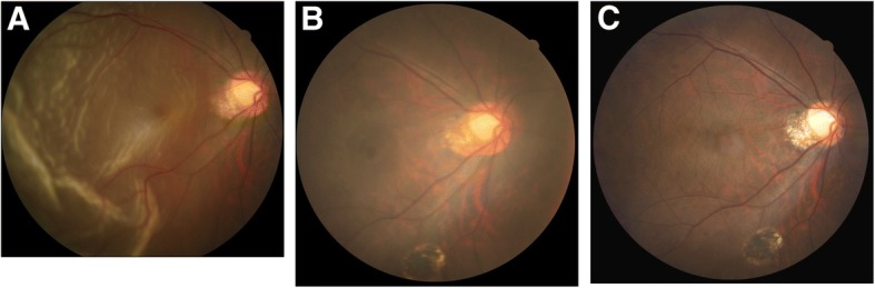
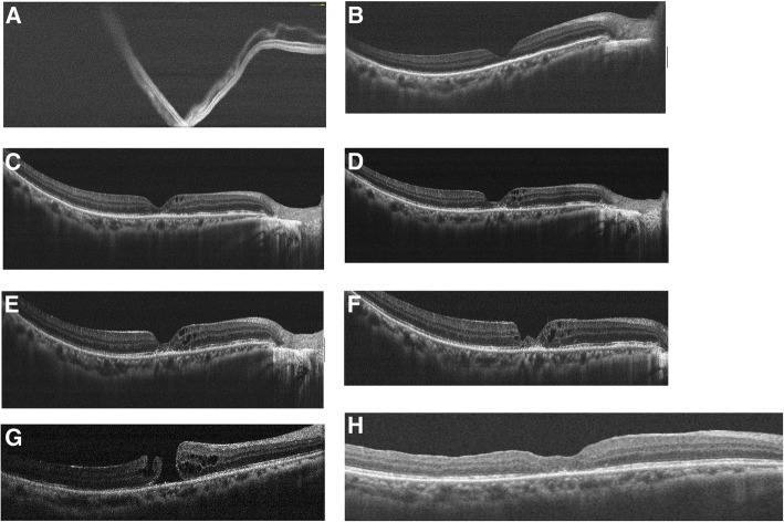
A 47-year-old female underwent 23G vitrectomy surgery to repair the macula-detached RRD successfully. However, intraretinal cysts initially developed two months after surgery. Cysts gradually increased in number and size, and cystoid macular edema was noted at the 5th month. Thereafter, inner retina dehiscence and a lamellar macular hole developed. The lamellar hole further dehisced and progressed into a full-thickness MH at the 10th month. The patient then received 23G vitrectomy and internal limiting membrane peeling surgery. OCT and fundus picture showed macular hole sealed 10 days afterward.
The mechanism of secondary MH included tangential traction, cystoid degeneration of macula, and glial migration. The sequential OCT studies provide evidence to support the disease mechanism of cystoid degeneration of the macula.
为了证明孔源性视网膜脱离(RRD)玻璃体切除术后全层黄斑裂孔(MH)的发生,并利用光学相干断层扫描(OCT)研究其可能的发病机制。
一名47岁女性成功接受了23G玻璃体切除术以修复黄斑脱离的RRD。然而,术后两个月视网膜内囊肿最初形成。囊肿数量和大小逐渐增加,在第5个月时出现黄斑囊样水肿。此后,内层视网膜裂开并形成板层黄斑裂孔。板层裂孔进一步裂开并在第10个月发展为全层MH。该患者随后接受了23G玻璃体切除术和内界膜剥除术。术后10天,OCT和眼底照片显示黄斑裂孔封闭。
继发性MH的机制包括切线牵引、黄斑囊样变性和胶质细胞迁移。连续的OCT研究为黄斑囊样变性的发病机制提供了证据支持。