Department of Neuroscience, University of Connecticut Health Center, Farmington, Connecticut.
Department of Chemistry, University of Texas at El Paso, El Paso, Texas.
J Cell Physiol. 2019 Jun;234(6):8683-8697. doi: 10.1002/jcp.27528. Epub 2018 Oct 14.
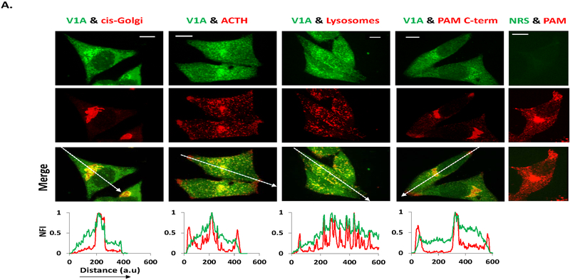
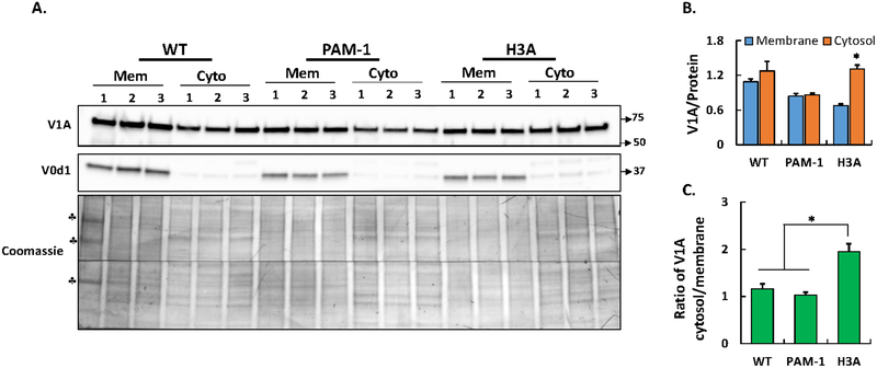
The biosynthetic and endocytic pathways of secretory cells are characterized by progressive luminal acidification, a process which is crucial for posttranslational modifications and membrane trafficking. This progressive fall in luminal pH is mainly achieved by the vacuolar-type-H ATPase (V-ATPase). V-ATPases are large, evolutionarily ancient rotary proton pumps that consist of a peripheral V1 complex, which hydrolyzes ATP, and an integral membrane V0 complex, which transports protons from the cytosol into the lumen. Upon sensing the desired luminal pH, V-ATPase activity is regulated by reversible dissociation of the complex into its V1 and V0 components. Molecular details of how intraluminal pH is sensed and transmitted to the cytosol are not fully understood. Peptidylglycine α-amidating mono-oxygenase (PAM; EC 1.14.17.3), a secretory pathway membrane enzyme which shares similar topology with two V-ATPase accessory proteins (Ac45 and prorenin receptor), has a pH-sensitive luminal linker region. Immunofluorescence and sucrose gradient analysis of peptidergic cells (AtT-20) identified distinct subcellular compartments exhibiting spatial co-occurrence of PAM and V-ATPase. In vitro binding assays demonstrated direct binding of the cytosolic domain of PAM to V1H. Blue native PAGE identified heterogeneous high-molecular weight complexes of PAM and V-ATPase. A PAM-1 mutant (PAM-1/H3A) with altered pH sensitivity had diminished ability to form high-molecular weight complexes. In addition, V-ATPase assembly status was altered in PAM-1/H3A expressing cells. Our analysis of the secretory and endocytic pathways of peptidergic cells supports the hypothesis that PAM serves as a luminal pH-sensor, regulating V-ATPase action by altering its assembly status.
分泌细胞的生物合成和内吞途径的特点是腔内腔逐渐酸化,这一过程对于翻译后修饰和膜运输至关重要。这种腔内 pH 的逐渐下降主要是通过液泡型 H+ATP 酶(V-ATPase)实现的。V-ATPases 是大型的、进化古老的旋转质子泵,由一个外周 V1 复合物组成,该复合物水解 ATP,以及一个完整的膜 V0 复合物,该复合物将质子从细胞质运送到腔内。当感应到所需的腔内 pH 值时,V-ATPase 活性通过复合物可逆地解离为其 V1 和 V0 组件来调节。腔内 pH 值如何被感应并传递到细胞质的分子细节尚不完全清楚。肽基甘氨酸 α-酰胺化单加氧酶(PAM;EC 1.14.17.3)是一种分泌途径膜酶,与两种 V-ATPase 辅助蛋白(Ac45 和前肾素受体)具有相似的拓扑结构,具有 pH 敏感的腔内连接区。肽能细胞(AtT-20)的免疫荧光和蔗糖梯度分析鉴定了具有 PAM 和 V-ATPase 空间共定位的不同亚细胞隔室。体外结合测定表明 PAM 的细胞质结构域与 V1H 直接结合。蓝色非变性 PAGE 鉴定了 PAM 和 V-ATPase 的异质高分子量复合物。具有改变的 pH 敏感性的 PAM-1 突变体(PAM-1/H3A)形成高分子量复合物的能力降低。此外,在表达 PAM-1/H3A 的细胞中 V-ATPase 组装状态发生改变。我们对肽能细胞的分泌和内吞途径的分析支持这样的假设,即 PAM 作为腔内 pH 传感器,通过改变其组装状态来调节 V-ATPase 的作用。