Department of Microbiology and Immunology, College of Medicine and Health Sciences, United Arab Emirates University, Al Ain, United Arab Emirates.
Department of Anatomy, College of Medicine and Health Sciences, United Arab Emirates University, Al Ain, United Arab Emirates.
Sci Rep. 2018 Oct 18;8(1):15438. doi: 10.1038/s41598-018-33758-4.
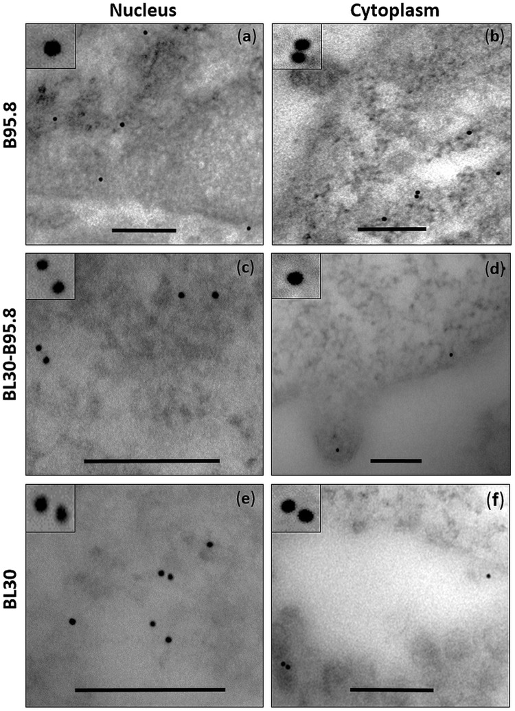
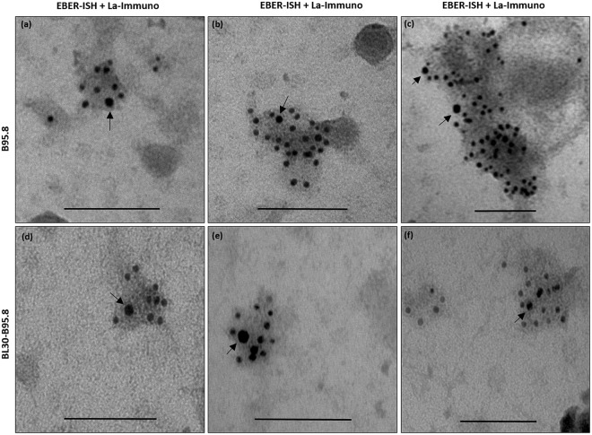
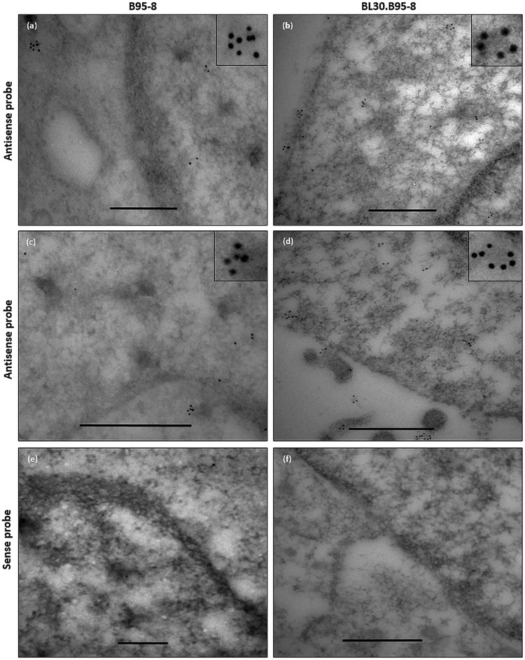
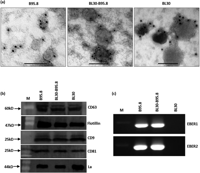
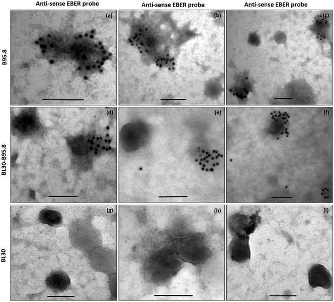
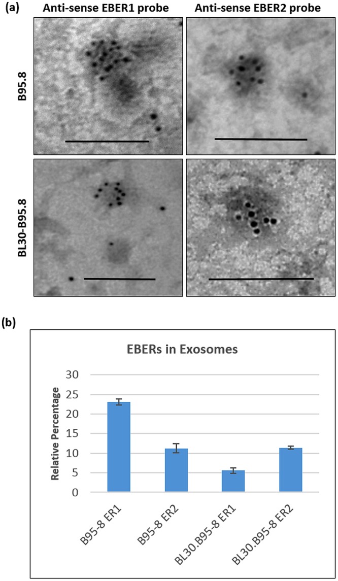
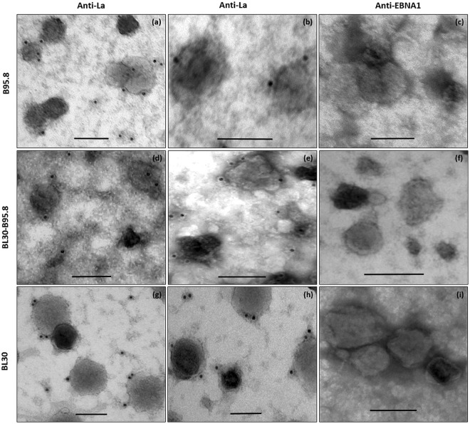
Epstein-Barr virus-encoded RNAs (EBER1 and EBER2) are two highly abundant, non-protein coding RNAs consistently expressed in all EBV infected cells, but their function remains poorly understood. Conventional in situ hybridization studies have indicated that these RNAs are present exclusively in the nucleus. We have recently demonstrated that EBERs can be excreted from infected cells via exosomes. However, the details of the steps involved in their excretion remain unknown. In this study, we aimed to directly track the journey of EBERs from the nucleus to the excretory exosomes of EBV immortalized B-lymphocytes. Using a combination of molecular and novel immuno-gold labelled electron microscopy (EM) based techniques, we demonstrate the presence of EBERs, not only in the nucleus, but also in the cytoplasm of EBV infected B cell lines. EBERs were also seen in exosomes shed from infected cells along with the EBER binding protein La. Our results show, for the first time, that at least a proportion of EBERs are transported from the nucleus to the cytoplasm where they appear to be loaded into multi-vesicular bodies for eventual excretion via exosomes.
EB 病毒编码的 RNA(EBER1 和 EBER2)是两种高度丰富的非编码 RNA,在所有感染 EBV 的细胞中持续表达,但它们的功能仍知之甚少。传统的原位杂交研究表明,这些 RNA 仅存在于细胞核中。我们最近证明,EBER 可以通过外泌体从感染的细胞中排出。然而,其排泄过程中涉及的具体步骤仍不清楚。在这项研究中,我们旨在直接追踪 EBER 从细胞核到 EBV 永生化 B 淋巴细胞的分泌性外泌体的旅程。我们使用分子和新型免疫金标记电子显微镜(EM)技术的组合,证明了 EBER 不仅存在于细胞核中,也存在于 EBV 感染的 B 细胞系的细胞质中。在感染细胞分泌的外泌体中也观察到了 EBER 以及与其结合的蛋白 La。我们的结果首次表明,至少一部分 EBER 从细胞核运输到细胞质,在那里它们似乎被装入多泡体,最终通过外泌体排出。