• 文献检索
  • 文档翻译
  • 深度研究
  • 学术资讯
  • Suppr Zotero 插件Zotero 插件
  • 邀请有礼
  • 套餐&价格
  • 历史记录
应用&插件
Suppr Zotero 插件Zotero 插件浏览器插件Mac 客户端Windows 客户端微信小程序
定价
高级版会员购买积分包购买API积分包
服务
文献检索文档翻译深度研究API 文档MCP 服务
关于我们
关于 Suppr公司介绍联系我们用户协议隐私条款
关注我们

Suppr 超能文献

核心技术专利:CN118964589B侵权必究
粤ICP备2023148730 号-1Suppr @ 2026

文献检索

告别复杂PubMed语法,用中文像聊天一样搜索,搜遍4000万医学文献。AI智能推荐,让科研检索更轻松。

立即免费搜索

文件翻译

保留排版,准确专业,支持PDF/Word/PPT等文件格式,支持 12+语言互译。

免费翻译文档

深度研究

AI帮你快速写综述,25分钟生成高质量综述,智能提取关键信息,辅助科研写作。

立即免费体验

房间隔的演化与发育

Evolution and Development of the Atrial Septum.

机构信息

Department of Medical Biology, Academic Medical Center, University of Amsterdam, The Netherlands.

Department of Bioscience, Zoophysiology, Aarhus University, Aarhus, Denmark.

出版信息

Anat Rec (Hoboken). 2019 Jan;302(1):32-48. doi: 10.1002/ar.23914. Epub 2018 Oct 18.

DOI:10.1002/ar.23914
PMID:30338646
原文链接:https://pmc.ncbi.nlm.nih.gov/articles/PMC6588001/
Abstract

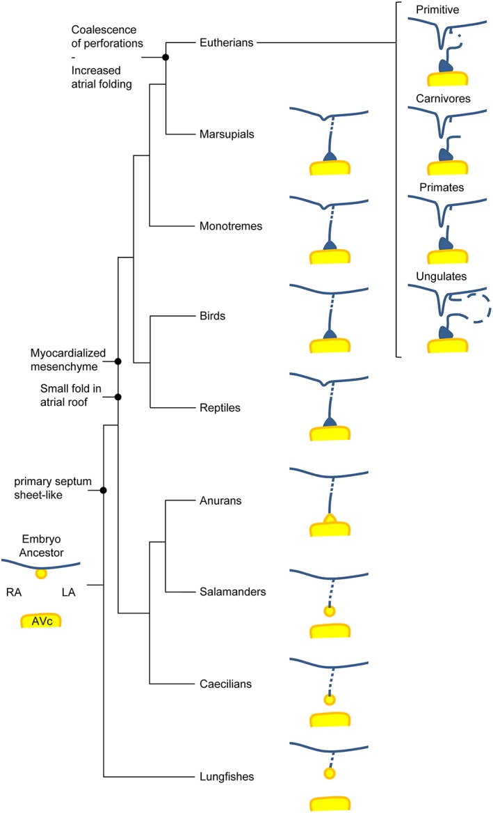
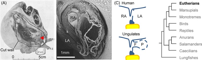
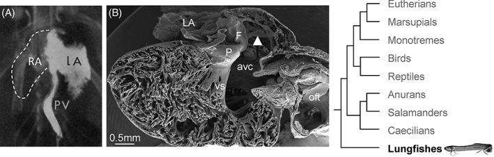
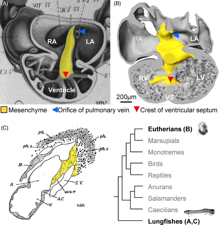
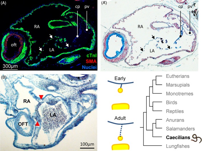
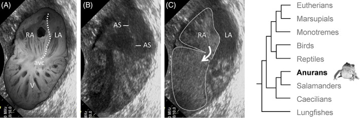
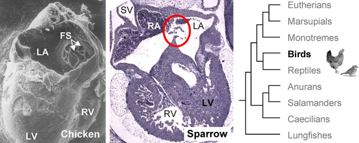
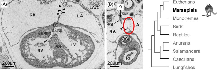
The complete division of the atrial cavity by a septum, resulting in a left and right atrium, is found in many amphibians and all amniotes (reptiles, birds, and mammals). Surprisingly, it is only in eutherian, or placental, mammals that full atrial septation necessitates addition from a second septum. The high incidence of incomplete closure of the atrial septum in human, so-called probe patency, suggests this manner of closure is inefficient. We review the evolution and development of the atrial septum to understand the peculiar means of forming the atrial septum in eutherian mammals. The most primitive atrial septum is found in lungfishes and comprises a myocardial component with a mesenchymal cap on its leading edge, reminiscent to the primary atrial septum of embryonic mammals before closure of the primary foramen. In reptiles, birds, and mammals, the primary foramen is closed by the mesenchymal tissues of the atrioventricular cushions, the dorsal mesenchymal protrusion, and the mesenchymal cap. These tissues are also found in lungfishes. The closure of the primary foramen is preceded by the development of secondary perforations in the septal myocardium. In all amniotes, with the exception of eutherian mammals, the secondary perforations do not coalesce to a secondary foramen. Instead, the secondary perforations persist and are sealed by myocardial and endocardial growth after birth or hatching. We suggest that the error-prone secondary foramen allows large volumes of oxygen-rich blood to reach the cardiac left side, needed to sustain the growth of the extraordinary large offspring that characterizes eutherian mammals. Anat Rec, 302:32-48, 2019. © 2018 The Authors. The Anatomical Record published by Wiley Periodicals, Inc. on behalf of American Association of Anatomists.

摘要

心房腔完全由中隔分隔,形成左心房和右心房,这在许多两栖动物和所有羊膜动物(爬行动物、鸟类和哺乳动物)中都有发现。令人惊讶的是,只有在真哺乳类或胎盘哺乳动物中,完全的心房分隔才需要第二个中隔的加入。人类的房间隔不完全闭合的发生率很高,即所谓的探针通畅性,这表明这种闭合方式效率低下。我们回顾了房间隔的进化和发育,以了解真哺乳类动物形成房间隔的特殊方式。最原始的房间隔见于肺鱼,由心肌成分组成,前缘有一个间质帽,类似于胚胎哺乳动物初级房间隔闭合前的初级房间隔。在爬行动物、鸟类和哺乳动物中,初级房间隔由房室瓣垫的间质组织、背侧间质突起和间质帽闭合。这些组织也存在于肺鱼中。初级房间隔的关闭之前,是间隔心肌中的次级穿孔的发育。在所有羊膜动物中,除了真哺乳类动物外,次级穿孔不会融合成次级房间隔。相反,次级穿孔在出生或孵化后会持续存在,并通过心肌和心内膜的生长来封闭。我们认为,易出错的次级房间隔允许大量富含氧气的血液进入心脏左侧,这是维持特征为真哺乳类动物的巨大后代生长所必需的。解剖学记录,302:32-48, 2019. © 2018 作者。解剖学记录由 Wiley 期刊出版公司代表美国解剖学家协会出版。

https://cdn.ncbi.nlm.nih.gov/pmc/blobs/7af6/6588001/bfa218227e02/AR-302-32-g014.jpg
https://cdn.ncbi.nlm.nih.gov/pmc/blobs/7af6/6588001/3603235f5d78/AR-302-32-g001.jpg
https://cdn.ncbi.nlm.nih.gov/pmc/blobs/7af6/6588001/4729e86eefc3/AR-302-32-g002.jpg
https://cdn.ncbi.nlm.nih.gov/pmc/blobs/7af6/6588001/bfeaa0dd8ca9/AR-302-32-g003.jpg
https://cdn.ncbi.nlm.nih.gov/pmc/blobs/7af6/6588001/9739156f7101/AR-302-32-g004.jpg
https://cdn.ncbi.nlm.nih.gov/pmc/blobs/7af6/6588001/7db7110ffde0/AR-302-32-g005.jpg
https://cdn.ncbi.nlm.nih.gov/pmc/blobs/7af6/6588001/0a9cd57638f9/AR-302-32-g006.jpg
https://cdn.ncbi.nlm.nih.gov/pmc/blobs/7af6/6588001/cd1ec956d888/AR-302-32-g007.jpg
https://cdn.ncbi.nlm.nih.gov/pmc/blobs/7af6/6588001/616197c8f289/AR-302-32-g008.jpg
https://cdn.ncbi.nlm.nih.gov/pmc/blobs/7af6/6588001/e98a9bad07c2/AR-302-32-g009.jpg
https://cdn.ncbi.nlm.nih.gov/pmc/blobs/7af6/6588001/fc903c3b345a/AR-302-32-g010.jpg
https://cdn.ncbi.nlm.nih.gov/pmc/blobs/7af6/6588001/f1f7397fda59/AR-302-32-g011.jpg
https://cdn.ncbi.nlm.nih.gov/pmc/blobs/7af6/6588001/5c3790e7d307/AR-302-32-g012.jpg
https://cdn.ncbi.nlm.nih.gov/pmc/blobs/7af6/6588001/96c23807bc92/AR-302-32-g013.jpg
https://cdn.ncbi.nlm.nih.gov/pmc/blobs/7af6/6588001/bfa218227e02/AR-302-32-g014.jpg

相似文献

1
Evolution and Development of the Atrial Septum.房间隔的演化与发育
Anat Rec (Hoboken). 2019 Jan;302(1):32-48. doi: 10.1002/ar.23914. Epub 2018 Oct 18.
2
Development of the atrial septum in relation to postnatal anatomy and interatrial communications.房间隔发育与出生后解剖结构及房间隔交通的关系。
Heart. 2017 Mar;103(6):456-462. doi: 10.1136/heartjnl-2016-310660. Epub 2016 Dec 21.
3
Low incidence of atrial septal defects in nonmammalian vertebrates.非哺乳动物脊椎动物中心房间隔缺损的发生率较低。
Evol Dev. 2020 May;22(3):241-256. doi: 10.1111/ede.12322. Epub 2019 Oct 9.
4
Atrial development in the human heart: an immunohistochemical study with emphasis on the role of mesenchymal tissues.人类心脏的心房发育:一项着重于间充质组织作用的免疫组织化学研究。
Anat Rec. 2000 Jul 1;259(3):288-300. doi: 10.1002/1097-0185(20000701)259:3<288::AID-AR60>3.0.CO;2-D.
5
Clarifying the morphology of the ostium primum defect.明确原发孔缺损的形态。
J Anat. 2015 Mar;226(3):244-57. doi: 10.1111/joa.12272. Epub 2015 Feb 9.
6
The development of septation in the four-chambered heart.四腔心分隔的发育
Anat Rec (Hoboken). 2014 Aug;297(8):1414-29. doi: 10.1002/ar.22949. Epub 2014 May 27.
7
Extreme variation in the atrial septation of caecilians (Amphibia: Gymnophiona).蚓螈(两栖纲:蚓螈目)心房分隔的极端变异。
J Anat. 2015 Jan;226(1):1-12. doi: 10.1111/joa.12255. Epub 2014 Nov 14.
8
Remodeling of the Embryonic Interventricular Communication in Regard to the Description and Classification of Ventricular Septal Defects.胚胎室间隔交通重塑与室间隔缺损的描述和分类。
Anat Rec (Hoboken). 2019 Jan;302(1):19-31. doi: 10.1002/ar.24020. Epub 2018 Nov 29.
9
Convergent evolutionary reduction of atrial septation in lungless salamanders.无肺螈类心房分隔的趋同进化简化
J Anat. 2017 Jan;230(1):16-29. doi: 10.1111/joa.12535. Epub 2016 Aug 25.
10
Clinical anatomy of the atrial septum with reference to its developmental components.房间隔的临床解剖学及其发育组成部分
Clin Anat. 1999;12(5):362-74. doi: 10.1002/(SICI)1098-2353(1999)12:5<362::AID-CA6>3.0.CO;2-F.

引用本文的文献

1
Morphological variations of the interatrial septum and potential implications in equine cardiology.马心脏学中房间隔的形态学变异及潜在影响
Sci Rep. 2025 May 12;15(1):16500. doi: 10.1038/s41598-025-01387-3.
2
Developmental and Evolutionary Heart Adaptations Through Structure-Function Relationships.通过结构-功能关系实现的发育和进化心脏适应
J Cardiovasc Dev Dis. 2025 Feb 22;12(3):83. doi: 10.3390/jcdd12030083.
3
Echocardiographic evaluation of the right atrial size and function: Relevance for clinical practice.右心房大小和功能的超声心动图评估:对临床实践的意义。

本文引用的文献

1
Total evidence phylogeny and evolutionary timescale for Australian faunivorous marsupials (Dasyuromorphia).澳大利亚食虫有袋类动物(袋鼬目)的全证据系统发育和进化时间尺度。
BMC Evol Biol. 2017 Dec 4;17(1):240. doi: 10.1186/s12862-017-1090-0.
2
Comparative anatomy of neonates of the three major mammalian groups (monotremes, marsupials, placentals) and implications for the ancestral mammalian neonate morphotype.三大哺乳动物类群(单孔目、有袋目、胎盘类)新生儿的比较解剖学及其对原始哺乳动物新生儿形态型的影响。
J Anat. 2017 Dec;231(6):798-822. doi: 10.1111/joa.12689. Epub 2017 Sep 28.
3
Molecular conservation of marsupial and eutherian placentation and lactation.
Am Heart J Plus. 2023 Feb 15;27:100274. doi: 10.1016/j.ahjo.2023.100274. eCollection 2023 Mar.
4
Genetic Alterations of Transcription Factors and Signaling Molecules Involved in the Development of Congenital Heart Defects-A Narrative Review.参与先天性心脏病发生发展的转录因子和信号分子的基因改变——一篇叙述性综述
Children (Basel). 2023 Apr 29;10(5):812. doi: 10.3390/children10050812.
5
Anatomy of the heart with the highest heart rate.心脏解剖图与最大心率。
J Anat. 2022 Jul;241(1):173-190. doi: 10.1111/joa.13640. Epub 2022 Feb 6.
6
The Genetic Landscape of Patent Foramen Ovale: A Systematic Review.卵圆孔未闭的遗传学特征:系统综述。
Genes (Basel). 2021 Dec 6;12(12):1953. doi: 10.3390/genes12121953.
7
Morphological Study of Fossa Ovalis in Formalin-Fixed Human Hearts and Its Clinical Importance.卵圆窝的福尔马林固定人心标本形态学研究及其临床意义。
Medicina (Kaunas). 2021 Nov 16;57(11):1254. doi: 10.3390/medicina57111254.
8
Anuran development: A reinvestigation of the conus arteriosus and gill formation in Bufo bufo throughout metamorphosis using micro-CT.蛙类发育:利用 micro-CT 对变态期Bufo bufo 的动脉圆锥和鳃形成进行重新研究
Anat Rec (Hoboken). 2022 May;305(5):1100-1111. doi: 10.1002/ar.24766. Epub 2021 Sep 27.
9
Role of the Epicardium in the Development of the Atrioventricular Valves and Its Relevance to the Pathogenesis of Myxomatous Valve Disease.心外膜在房室瓣发育中的作用及其与黏液瘤样瓣膜病发病机制的相关性。
J Cardiovasc Dev Dis. 2021 May 12;8(5):54. doi: 10.3390/jcdd8050054.
10
The Mesenchymal Cap of the Atrial Septum and Atrial and Atrioventricular Septation.房间隔的间充质帽与心房及房室间隔形成
J Cardiovasc Dev Dis. 2020 Nov 4;7(4):50. doi: 10.3390/jcdd7040050.
有袋类动物和真兽类动物胎盘和哺乳的分子保守性。
Elife. 2017 Sep 12;6:e27450. doi: 10.7554/eLife.27450.
4
Sequential segmental analysis of the crocodilian heart.鳄鱼心脏的连续节段分析
J Anat. 2017 Oct;231(4):484-499. doi: 10.1111/joa.12661. Epub 2017 Aug 1.
5
Morpho-functional characterization of the systemic venous pole of the reptile heart.爬行动物心脏体静脉干的形态功能特征。
Sci Rep. 2017 Jul 27;7(1):6644. doi: 10.1038/s41598-017-06291-z.
6
Editorial Commentary: Closure of the patent foramen ovale viewed from a different angle.编辑评论:从不同角度看待卵圆孔未闭的封堵
Trends Cardiovasc Med. 2017 Nov;27(8):582-584. doi: 10.1016/j.tcm.2017.07.003. Epub 2017 Jul 11.
7
Patent foramen ovale and cryptogenic stroke.卵圆孔未闭与隐源性卒中。
Trends Cardiovasc Med. 2017 Nov;27(8):575-581. doi: 10.1016/j.tcm.2017.06.016. Epub 2017 Jul 4.
8
Outflow tract septation and the aortic arch system in reptiles: lessons for understanding the mammalian heart.爬行动物的流出道分隔与主动脉弓系统:对理解哺乳动物心脏的启示
Evodevo. 2017 May 10;8:9. doi: 10.1186/s13227-017-0072-z. eCollection 2017.
9
The World Health Organization Fetal Growth Charts: A Multinational Longitudinal Study of Ultrasound Biometric Measurements and Estimated Fetal Weight.世界卫生组织胎儿生长图表:超声生物测量与估计胎儿体重的多国纵向研究
PLoS Med. 2017 Jan 24;14(1):e1002220. doi: 10.1371/journal.pmed.1002220. eCollection 2017 Jan.
10
Development of the atrial septum in relation to postnatal anatomy and interatrial communications.房间隔发育与出生后解剖结构及房间隔交通的关系。
Heart. 2017 Mar;103(6):456-462. doi: 10.1136/heartjnl-2016-310660. Epub 2016 Dec 21.