Ardani I Gusti Aju Wahju, Susanti Bintiana, Djaharu'ddin Irwadi
Department of Orthodontics, Faculty of Dental Medicine, Universitas Airlangga, Surabaya, Indonesia,
Department of Orthodontics, Faculty of Dental Medicine, Universitas Airlangga, Surabaya, Indonesia.
Clin Cosmet Investig Dent. 2018 Oct 11;10:211-220. doi: 10.2147/CCIDE.S173648. eCollection 2018.
Two types of orthodontic elastics exist based on their material, latex and nonlatex, each of which has different properties in clinical use. Some of the differences include their initial force and force degradation over time. This study was conducted to compare the force changes in both materials.
To evaluate the force degradation of latex and nonlatex elastics under moderate stretching over time.
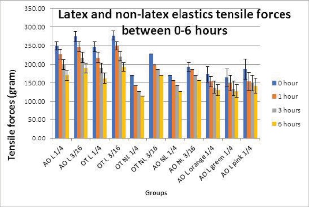
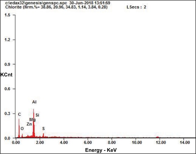
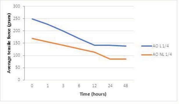
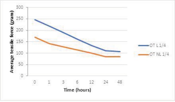
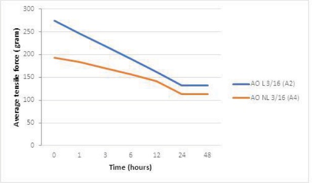
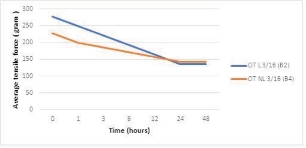
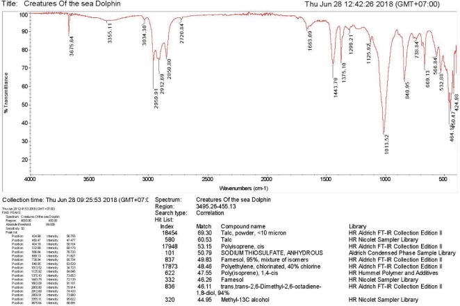
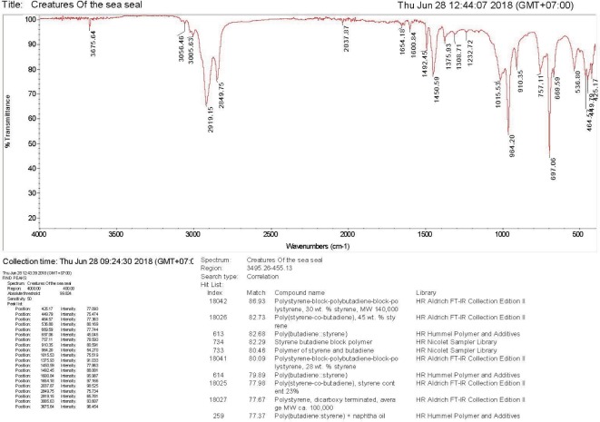
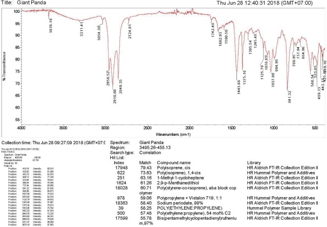
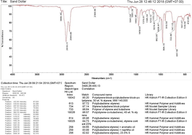
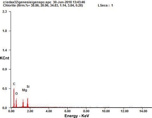
Medium-force orthodontic latex and nonlatex elastics from American Orthodontics (AO) and Ortho Technology (OT) of lumen size 1/4 inches and 3/16 inches (total sample 110 elastics) were submerged in artificial saliva (pH 6.7) and incubated for 48 hours. Then, the elastic force was measured at the following time intervals: initial, 1, 3, 6, 12, 24, and 48 hours. Orthodontic latex and nonlatex elastics from AO and OT were analyzed using Fourier-Transform Infrared Spectroscopy and energy-dispersive X-ray to know the chemical bond structure and elements.
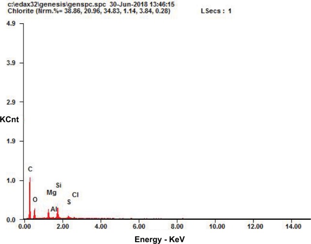
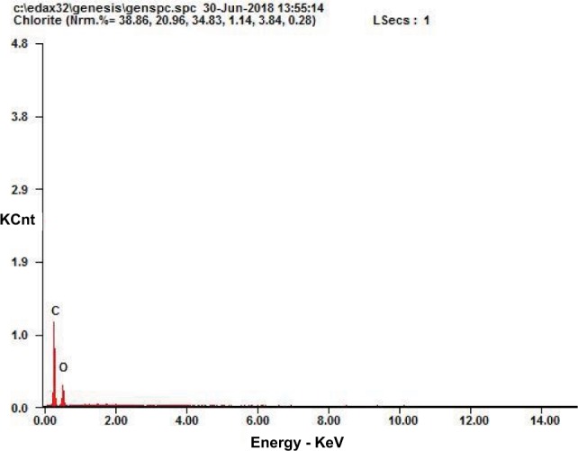
There was a statistically significant difference between latex and nonlatex force degradation over a period of 0-24 hours (<0.05), while no significant difference existed between 24-48 hours (>0.05). Force degradation of latex elastics was higher than that of nonlatex elastics. Energy-dispersive X-ray results on nonlatex elastic bands from OT and AO showed higher C element in the latex elastic band from OT, while the latex elastic band from AO had higher Al element.
Medium-force latex and nonlatex elastics 1/4 inches and 3/16 inches in size both showed force degradation at 1, 3, 6, 12, and 24-hour intervals under 30 mm stretching when kept in artificial saliva (pH 6.7) and incubated at 37°C for 48 hours.
正畸弹力圈根据其材料分为两种类型,即乳胶类和非乳胶类,它们在临床使用中具有不同的特性。其中一些差异包括它们的初始力以及力随时间的衰减情况。本研究旨在比较这两种材料的力变化情况。
评估乳胶和非乳胶弹力圈在适度拉伸下随时间的力衰减情况。
将美国正畸公司(AO)和正畸技术公司(OT)生产的内径为1/4英寸和3/16英寸的中等力量正畸乳胶和非乳胶弹力圈(共110个弹力圈样本)浸泡在人工唾液(pH值6.7)中,并孵育48小时。然后,在以下时间间隔测量弹力:初始、1小时、3小时、6小时、12小时、24小时和48小时。使用傅里叶变换红外光谱和能量色散X射线对AO和OT的正畸乳胶和非乳胶弹力圈进行分析,以了解化学键结构和元素。
在0至24小时内,乳胶和非乳胶弹力圈的力衰减存在统计学显著差异(<0.05),而在24至48小时之间则无显著差异(>0.05)。乳胶弹力圈的力衰减高于非乳胶弹力圈。OT和AO的非乳胶弹力带的能量色散X射线结果显示,OT的乳胶弹力带中的C元素含量较高,而AO的乳胶弹力带中的Al元素含量较高。
内径为1/4英寸和3/16英寸的中等力量乳胶和非乳胶弹力圈在30毫米拉伸下,于人工唾液(pH值6.7)中在37°C孵育48小时后,在1小时、3小时、6小时、12小时和24小时的时间间隔均表现出力衰减。