Department of Neuroscience, Trafford Centre for Biomedical Research, Brighton and Sussex Medical School, Brighton, UK
Brighton and Sussex University Hospitals NHS Trust, Princess Royal Hospital, Haywards Heath, UK.
J Neurol Neurosurg Psychiatry. 2019 Apr;90(4):404-411. doi: 10.1136/jnnp-2018-318830. Epub 2018 Oct 25.
Corticospinal tract (CST) degeneration and cortical atrophy are consistent features of amyotrophic lateral sclerosis (ALS). We hypothesised that neurite orientation dispersion and density imaging (NODDI), a multicompartment model of diffusion MRI, would reveal microstructural changes associated with ALS within the CST and precentral gyrus (PCG) 'in vivo'.
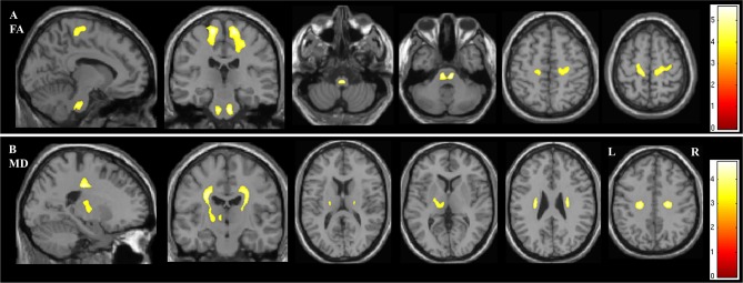
23 participants with sporadic ALS and 23 healthy controls underwent diffusion MRI. Neurite density index (NDI), orientation dispersion index (ODI) and free water fraction (isotropic compartment (ISO)) were derived. Whole brain voxel-wise analysis was performed to assess for group differences. Standard diffusion tensor imaging (DTI) parameters were computed for comparison. Subgroup analysis was performed to investigate for NODDI parameter differences relating to bulbar involvement. Correlation of NODDI parameters with clinical variables were also explored. The results were accepted as significant where p<0.05 after family-wise error correction at the cluster level, clusters formed with p<0.001.
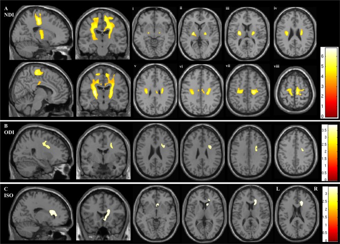
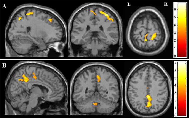
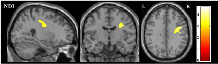
In the ALS group NDI was reduced in the extensive regions of the CST, the corpus callosum and the right PCG. ODI was reduced in the right anterior internal capsule and the right PCG. Significant differences in NDI were detected between subgroups stratified according to the presence or absence of bulbar involvement. ODI and ISO correlated with disease duration.
NODDI demonstrates that axonal loss within the CST is a core feature of degeneration in ALS. This is the main factor contributing to the altered diffusivity profile detected using DTI. NODDI also identified dendritic alterations within the PCG, suggesting microstructural cortical dendritic changes occur together with CST axonal damage.
皮质脊髓束(CST)退变和皮质萎缩是肌萎缩侧索硬化症(ALS)的一致特征。我们假设,神经丝取向分散和密度成像(NODDI),一种扩散 MRI 的多室模型,将揭示与 ALS 相关的 CST 和中央前回(PCG)内的微观结构变化“体内”。
23 名散发性 ALS 患者和 23 名健康对照者接受了扩散 MRI 检查。得出神经丝密度指数(NDI)、取向分散指数(ODI)和自由水分数(各向同性室(ISO))。进行全脑体素分析以评估组间差异。计算了标准扩散张量成像(DTI)参数进行比较。进行了亚组分析,以研究与球部受累相关的 NODDI 参数差异。还探讨了 NODDI 参数与临床变量的相关性。在簇级进行了家族性错误校正后,p<0.05 被认为具有统计学意义,簇的形成 p<0.001。
在 ALS 组中,CST 的广泛区域、胼胝体和右侧 PCG 的 NDI 降低。右侧前内囊和右侧 PCG 的 ODI 降低。根据有无球部受累进行分层的亚组之间,NDI 存在显著差异。ODI 和 ISO 与疾病持续时间相关。
NODDI 表明 CST 内的轴突丢失是 ALS 变性的核心特征。这是使用 DTI 检测到的扩散率谱改变的主要因素。NODDI 还在 PCG 中发现了树突改变,表明皮质树突微结构变化与 CST 轴突损伤同时发生。