Aktaa Suleman, Fatania Kavi, Gains Claire, White Hazel
Department of Cardiology, Mid Yorkshire Hospitals NHS Trust, Wakefield, West Yorkshire, UK.
Department of Radiology, Mid Yorkshire Hospitals NHS Trust, Wakefield, West Yorkshire, UK.
BMJ Case Rep. 2018 Oct 25;2018:bcr-2018-226318. doi: 10.1136/bcr-2018-226318.
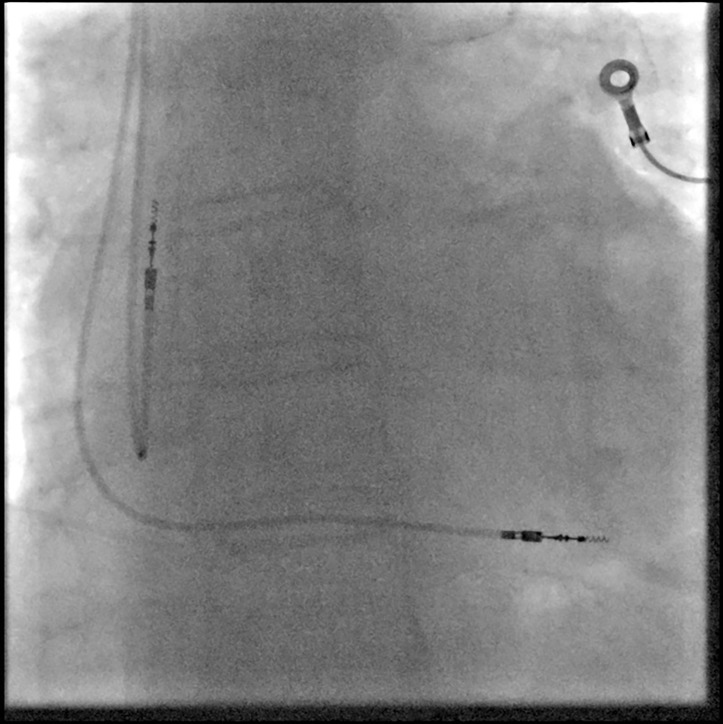
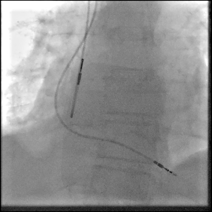
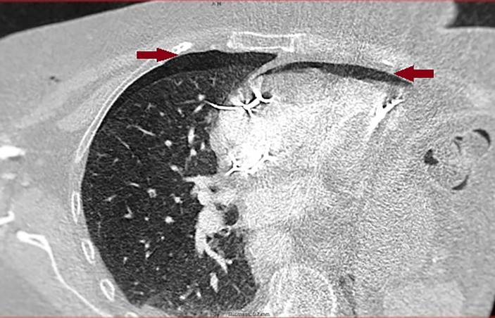
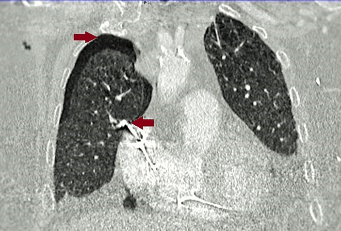
Permanent pacemaker (PPM) implantation is an increasingly common procedure with complication rate estimated between 3% and 6%. Cardiac perforation by pacemaker lead(s) is rare, but a previous study has shown that it is probably an underdiagnosed complication. We are presenting a case of a patient who presented 5 days after PPM insertion with new-onset pleuritic chest pain. She had a normal chest X-ray (CXR), and acceptable pacing checks. However, a CT scan of the chest showed pneumopericardium and pneumothorax secondary to atrial lead perforation. The pain only settled by replacing the atrial lead. A repeat chest CT scan a few months later showed complete resolution of the pneumopericardium and pneumothorax. We believe that cardiac perforation can be easily missed if associated with normal CXR and acceptable pacing parameters. Unexplained chest pain following PPM insertion might be the only clue for such complication, although it might not always be present.
植入永久性起搏器(PPM)是一种越来越常见的手术,其并发症发生率估计在3%至6%之间。起搏器导线导致的心脏穿孔很少见,但先前的一项研究表明,这可能是一种诊断不足的并发症。我们报告一例患者,在植入PPM 5天后出现新发胸膜炎性胸痛。她的胸部X光片(CXR)正常,起搏检查结果也可接受。然而,胸部CT扫描显示心房导线穿孔继发心包积气和气胸。通过更换心房导线,疼痛才得以缓解。几个月后重复进行的胸部CT扫描显示心包积气和气胸完全消失。我们认为,如果心脏穿孔伴有正常的CXR和可接受的起搏参数,很容易被漏诊。PPM植入后不明原因的胸痛可能是这种并发症的唯一线索,尽管并非总是出现。