Department of Ophthalmology and Visual Sciences, Vanderbilt Eye Institute, Vanderbilt University Medical Center, Nashville, TN, 37232, USA.
Department of Medicine, Vanderbilt University Medical Center, Nashville, TN, 37232, USA.
Cell Death Dis. 2018 Oct 26;9(11):1097. doi: 10.1038/s41419-018-1061-4.
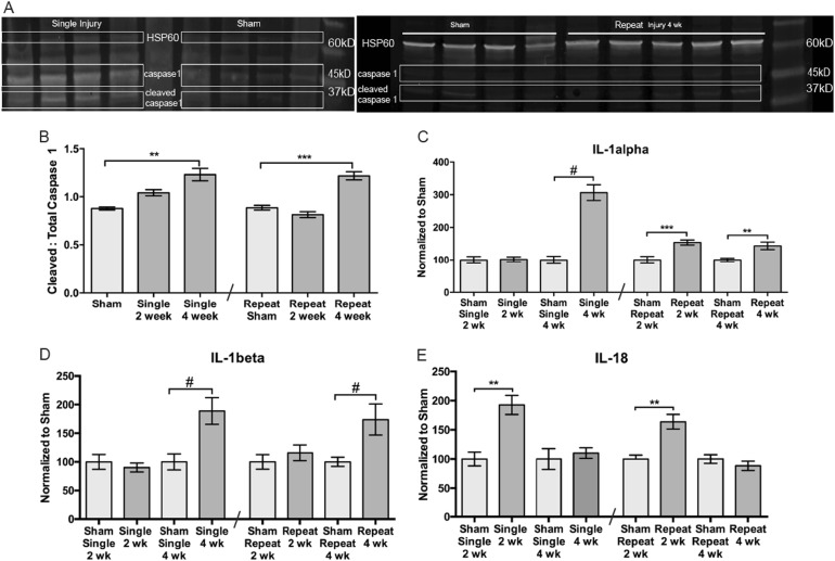
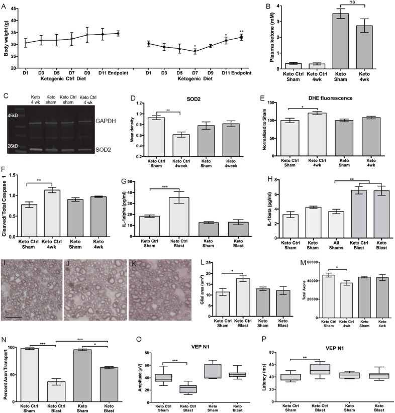
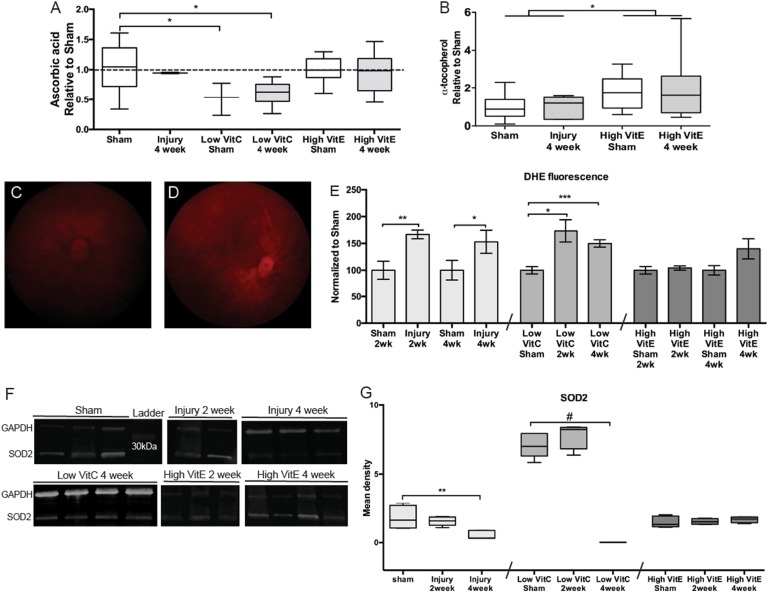
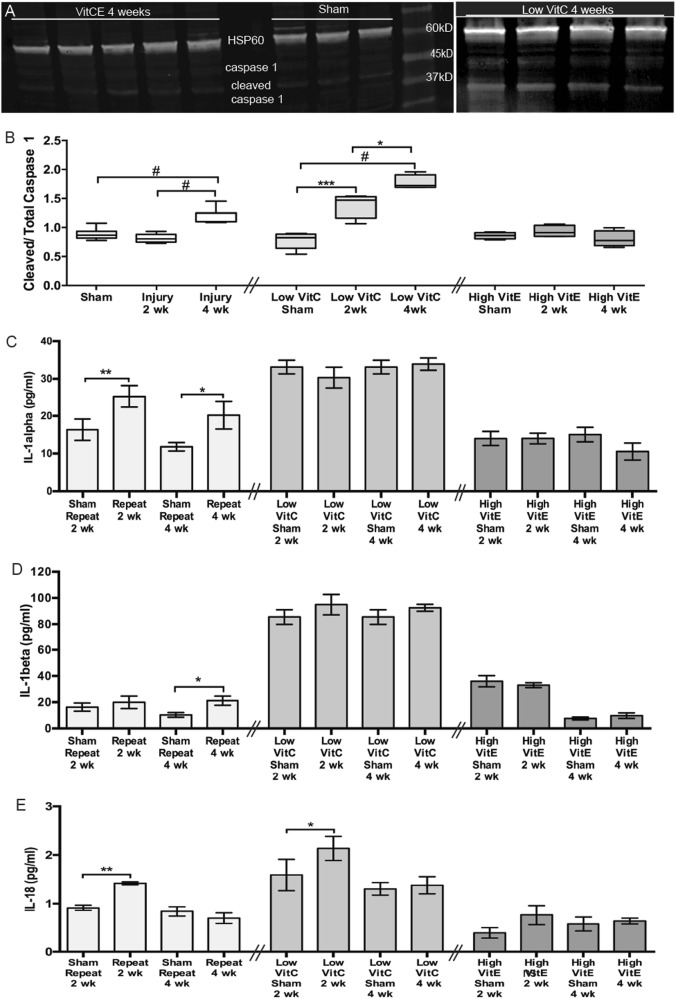
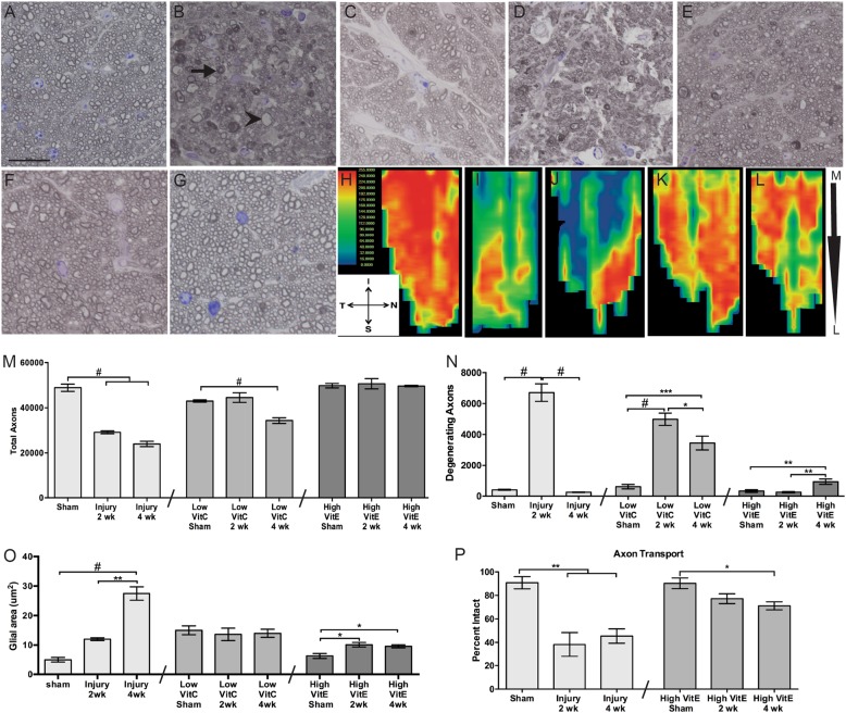
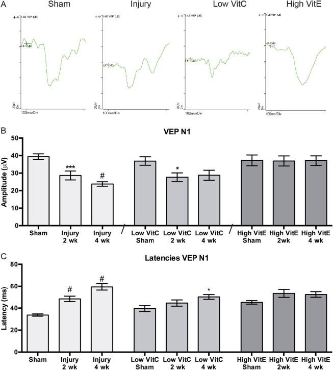
We investigated the role of oxidative stress and the inflammasome in trauma-induced axon degeneration and vision loss using a mouse model. The left eyes of male mice were exposed to over-pressure air waves. Wild-type C57Bl/6 mice were fed normal, high-vitamin-E (VitE), ketogenic or ketogenic-control diets. Mice lacking the ability to produce vitamin C (VitC) were maintained on a low-VitC diet. Visual evoked potentials (VEPs) and retinal superoxide levels were measured in vivo. Tissue was collected for biochemical and histological analysis. Injury increased retinal superoxide, decreased SOD2, and increased cleaved caspase-1, IL-1α, IL-1β, and IL-18 levels. Low-VitC exacerbated the changes and the high-VitE diet mitigated them, suggesting that oxidative stress led to the increase in IL-1α and activation of the inflammasome. The injury caused loss of nearly 50% of optic nerve axons at 2 weeks and astrocyte hypertrophy in mice on normal diet, both of which were prevented by the high-VitE diet. The VEP amplitude was decreased after injury in both control-diet and low-VitC mice, but not in the high-VitE-diet mice. The ketogenic diet also prevented the increase in superoxide levels and IL-1α, but had no effect on IL-1β. Despite this, the ketogenic diet preserved optic nerve axons, prevented astrocyte hypertrophy, and preserved the VEP amplitude. These data suggest that oxidative stress induces priming and activation of the inflammasome pathway after neurotrauma of the visual system. Further, blocking the activation of the inflammasome pathway may be an effective post-injury intervention.
我们使用小鼠模型研究了氧化应激和炎性小体在创伤性轴突变性和视力丧失中的作用。雄性小鼠的左眼暴露于超压气浪中。野生型 C57Bl/6 小鼠喂食正常、高维生素 E(VitE)、生酮或生酮对照饮食。缺乏产生维生素 C(VitC)能力的小鼠维持在低 VitC 饮食。在体内测量视觉诱发电位(VEPs)和视网膜超氧化物水平。收集组织进行生化和组织学分析。损伤增加了视网膜超氧化物,降低了 SOD2,并增加了切割的 caspase-1、IL-1α、IL-1β 和 IL-18 水平。低 VitC 加重了这些变化,而高 VitE 饮食减轻了这些变化,表明氧化应激导致了 IL-1α 的增加和炎性小体的激活。损伤导致正常饮食小鼠视神经轴突丢失近 50%,2 周后星形胶质细胞肥大,高 VitE 饮食可预防这些变化。损伤后,对照饮食和低 VitC 饮食小鼠的 VEP 幅度均降低,但高 VitE 饮食小鼠的 VEP 幅度未降低。生酮饮食也可预防超氧化物水平和 IL-1α 的增加,但对 IL-1β 没有影响。尽管如此,生酮饮食仍可保留视神经轴突,防止星形胶质细胞肥大,维持 VEP 幅度。这些数据表明,氧化应激在视觉系统神经损伤后诱导炎性小体途径的启动和激活。进一步,阻断炎性小体途径的激活可能是一种有效的损伤后干预措施。