Division of Otolaryngology-Head and Neck Surgery, University of Alberta, 1E4 Walter Mackenzie Center, 8440 112 Street, Edmonton, AB, T6G 2B7, Canada.
Department of Radiology & Diagnostic Imaging, Royal Alexandra Hospital, 1046 Royal Alexandra Hospital - Diagnostic Treatment Center, 2J2.00 WC Mackenzie Health Sciences Centre, 8440 112 Street, Edmonton, AB, T6G 2R7, Canada.
J Otolaryngol Head Neck Surg. 2018 Oct 26;47(1):65. doi: 10.1186/s40463-018-0307-6.
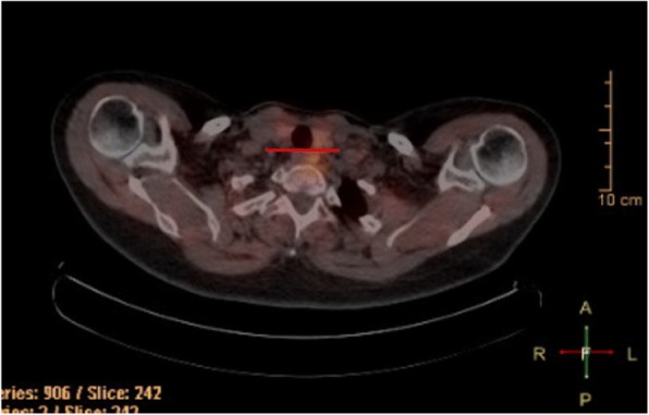
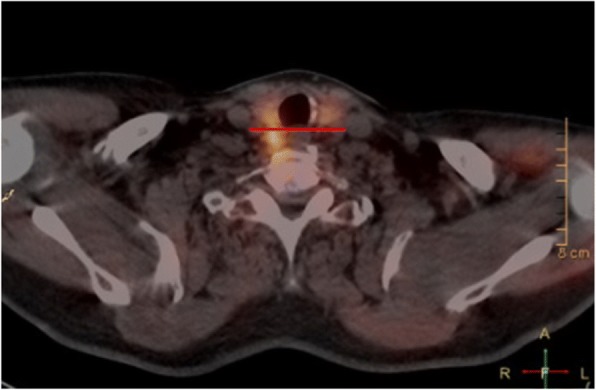
Accurate localization of parathyroid adenomas is of critical importance in surgical planning for minimally invasive parathyroidectomy. SPECT/CT is considered the investigation of choice but has limitations regarding localization of superior versus inferior adenomas. We proposed a novel method for localization using SPECT/CT by determining the anterior-posterior relationship of the adenoma to a horizontal line in the coronal plane through the tracheoesophageal groove. Our objective was to determine the accuracy, validity, and inter-rater reliability of this method.
This was a retrospective review of patients who underwent parathyroidectomy for a single adenoma between 2010-2017. SPECT/CT images were reviewed by two staff Otolaryngologists, a Radiologist, an Otolaryngology fellow and Otolaryngology resident. Results were compared using intra-operative report as the gold standard. Overall accuracy in determining superior/inferior and right/left adenomas was calculated, as well as Cohen's Kappa to determine agreement with operative report and inter-rater reliability. The performance was compared to that of the original radiology report.
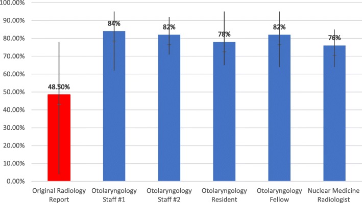
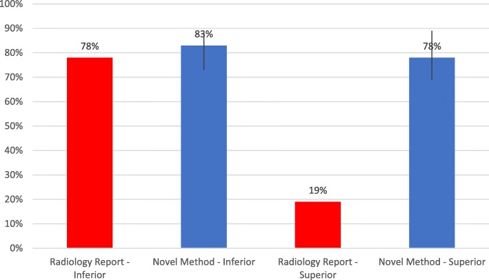
One hundred thirty patients met criteria and were included. Our method correctly identified the location of the adenoma in terms of both side and superior/inferior position in 80.4% [76 - 84%] of patients, which considerably outperformed the original radiology report at 48.5% [4-78%] accuracy. The agreement level between our method and operative report was high (Kappa=0.717 [0.691-0.743]), as was the inter-rater reliability (Kappa=0.706 [0.674-0.738]).
We report a novel method for localization of parathyroid adenomas using SPECT/CT which outperforms standard radiology reporting. This tool can be used by surgeons and radiologists to better inform and plan for minimally invasive parathyroidectomy.
甲状旁腺腺瘤的准确定位对微创甲状旁腺切除术的手术规划至关重要。SPECT/CT 被认为是首选的检查方法,但在定位上腺与下腺腺瘤方面存在局限性。我们提出了一种使用 SPECT/CT 进行定位的新方法,通过确定腺瘤在冠状面经气管食管沟的水平线的前后关系来实现。我们的目的是确定该方法的准确性、有效性和组内一致性。
这是一项回顾性研究,纳入了 2010 年至 2017 年间因单发腺瘤而行甲状旁腺切除术的患者。由两名耳鼻喉科医生、一名放射科医生、一名耳鼻喉科住院医师和一名耳鼻喉科住院医师对 SPECT/CT 图像进行回顾。以术中报告为金标准比较结果。计算确定上/下和右/左侧腺瘤的总体准确性,并使用 Cohen's Kappa 确定与手术报告的一致性和组内一致性。将其与原始放射学报告进行比较。
符合标准的 130 例患者被纳入研究。我们的方法在 80.4%[76-84%]的患者中正确确定了腺瘤的位置,无论是在侧位还是在上/下位,明显优于原始放射学报告的 48.5%[4-78%]的准确性。我们的方法与手术报告之间的一致性水平较高(Kappa=0.717[0.691-0.743]),组内一致性也较高(Kappa=0.706[0.674-0.738])。
我们报告了一种使用 SPECT/CT 定位甲状旁腺腺瘤的新方法,该方法优于标准放射学报告。该工具可由外科医生和放射科医生使用,以便更好地为微创甲状旁腺切除术提供信息和规划。