1 Department of Endocrinology and Internal Medicine Aarhus University Hospital Aarhus Denmark.
4 Department of Internal Medicine Regional Hospital Horsens Horsens Denmark.
J Am Heart Assoc. 2018 Aug 21;7(16):e008677. doi: 10.1161/JAHA.118.008677.
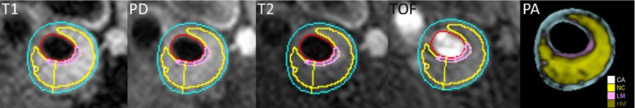
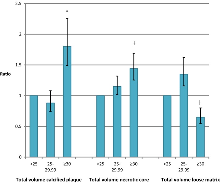
Background Ischemic stroke from carotid plaque embolism remains a major cause of morbidity in patients with type 2 diabetes mellitus (T2 DM ). However, the effect of early T2 DM and obesity on carotid remodeling and plaque burden remains elusive. We assessed carotid remodeling and plaque composition by carotid magnetic resonance imaging in patients with short-duration T2 DM compared with a sex- and age-matched control group. Methods and Results One hundred patients with T2 DM (duration <5 years) and 100 sex- and age-matched controls underwent bilateral carotid artery magnetic resonance imaging in a 1.5-T magnetic resonance imaging scanner. Plaque burden was quantified by normalized wall index, maximum wall thickness, maximum wall area, and minimum lumen size. Plaque morphology was quantified by calcified plaque volume, necrotic core volume, and loose matrix volume. Magnetic resonance imaging data were available for 149 and 177 carotid arteries from T2 DM patients and controls, respectively. Adjusted for age and sex, T2 DM was associated with increased plaque burden indicated by a higher normalized wall index (ratio 1.03 [95% confidence interval, 1.002; 1.06], P=0.03), and negative remodeling indicated by a lower minimum lumen area (ratio 0.81 [0.74; 0.89], P<0.001), and lower maximum wall area (ratio 0.94 [0.88; 1.00], P=0.048) compared with controls. In both T2 DM and controls, body mass index ≥30.0 kg/m was associated with an 80% increase in total calcified plaque volume, and a 44% increase in necrotic core volume compared with body mass index <25.0 kg/m. Conclusions Short-duration T2 DM was associated with increased carotid plaque burden and negative remodeling. Obesity was associated with increased carotid artery necrotic core volume and calcification independently of diabetes mellitus status. Clinical Trial Registration URL : https://www.clinicaltrials.gov . Unique identifier: NCT 00674271.
颈动脉斑块栓塞引起的缺血性脑卒中仍然是 2 型糖尿病(T2DM)患者发病率的主要原因。然而,早期 T2DM 和肥胖对颈动脉重构和斑块负担的影响仍不清楚。我们通过颈动脉磁共振成像评估了短期 T2DM 患者与性别和年龄匹配的对照组之间的颈动脉重构和斑块成分。
100 例 T2DM(病程<5 年)患者和 100 名性别和年龄匹配的对照组患者在 1.5-T 磁共振成像扫描仪上进行双侧颈动脉磁共振成像。通过标准化管壁指数、最大管壁厚度、最大管壁面积和最小管腔面积来量化斑块负担。通过钙化斑块体积、坏死核心体积和疏松基质体积来量化斑块形态。T2DM 患者和对照组的磁共振成像数据分别可用于 149 条和 177 条颈动脉。校正年龄和性别后,T2DM 与较高的斑块负担相关,表现为更高的标准化管壁指数(比值 1.03[95%置信区间,1.002;1.06],P=0.03),以及负性重构,表现为更小的最小管腔面积(比值 0.81[0.74;0.89],P<0.001)和更小的最大管壁面积(比值 0.94[0.88;1.00],P=0.048)。在 T2DM 和对照组中,体重指数(BMI)≥30.0kg/m2与总钙化斑块体积增加 80%和坏死核心体积增加 44%相关,而 BMI<25.0kg/m2。
短期 T2DM 与颈动脉斑块负担增加和负性重构有关。肥胖与颈动脉坏死核心体积和钙化的增加有关,与糖尿病状态无关。
https://www.clinicaltrials.gov。唯一标识符:NCT 00674271。