• 文献检索
  • 文档翻译
  • 深度研究
  • 学术资讯
  • Suppr Zotero 插件Zotero 插件
  • 邀请有礼
  • 套餐&价格
  • 历史记录
应用&插件
Suppr Zotero 插件Zotero 插件浏览器插件Mac 客户端Windows 客户端微信小程序
定价
高级版会员购买积分包购买API积分包
服务
文献检索文档翻译深度研究API 文档MCP 服务
关于我们
关于 Suppr公司介绍联系我们用户协议隐私条款
关注我们

Suppr 超能文献

核心技术专利:CN118964589B侵权必究
粤ICP备2023148730 号-1Suppr @ 2026

文献检索

告别复杂PubMed语法,用中文像聊天一样搜索,搜遍4000万医学文献。AI智能推荐,让科研检索更轻松。

立即免费搜索

文件翻译

保留排版,准确专业,支持PDF/Word/PPT等文件格式,支持 12+语言互译。

免费翻译文档

深度研究

AI帮你快速写综述,25分钟生成高质量综述,智能提取关键信息,辅助科研写作。

立即免费体验

单细胞水平下用于细胞力学特性表征的光镊技术综述

A Comprehensive Review of Optical Stretcher for Cell Mechanical Characterization at Single-Cell Level.

作者信息

Yang Tie, Bragheri Francesca, Minzioni Paolo

机构信息

Department of Electrical, Computer, and Biomedical Engineering, Università di Pavia, Via Ferrata 5A, Pavia 27100, Italy.

Institute of Photonics and Nanotechnology, CNR & Department of Physics, Politecnico di Milano, Piazza Leonardo da Vinci 32, Milano 20133, Italy.

出版信息

Micromachines (Basel). 2016 May 13;7(5):90. doi: 10.3390/mi7050090.

DOI:10.3390/mi7050090
PMID:30404265
原文链接:https://pmc.ncbi.nlm.nih.gov/articles/PMC6189960/
Abstract

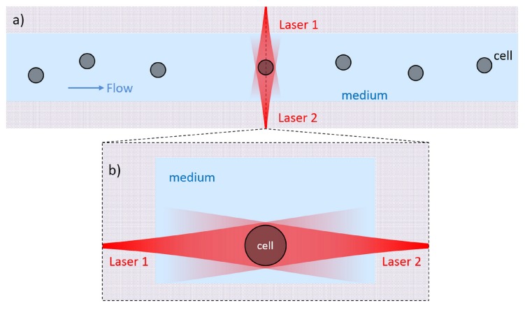
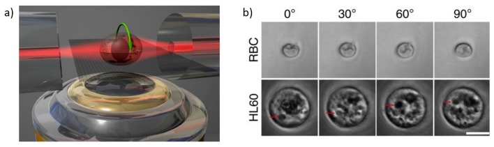
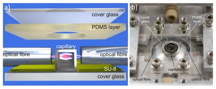
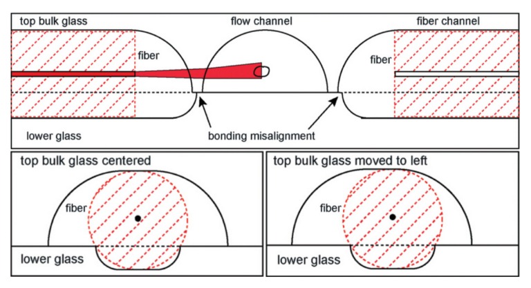
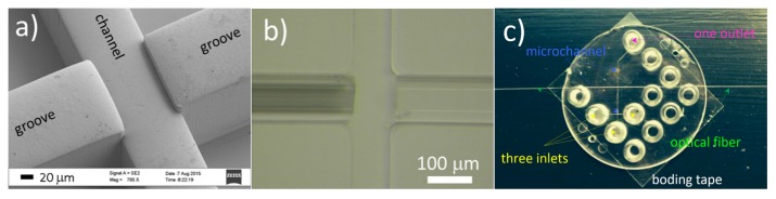
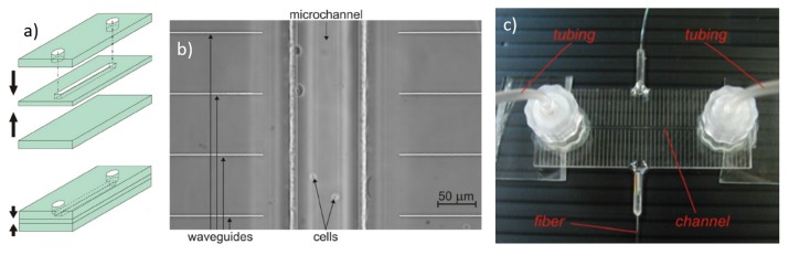
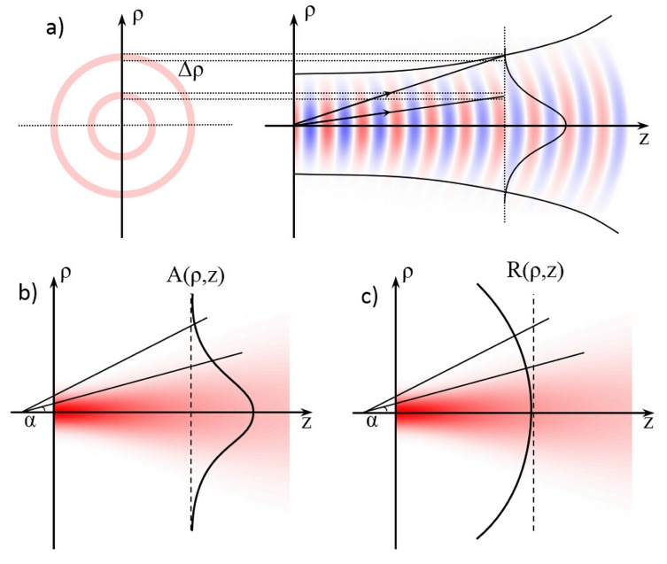
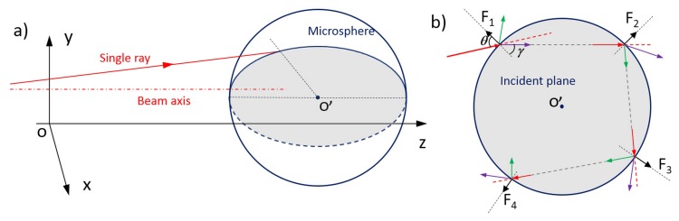
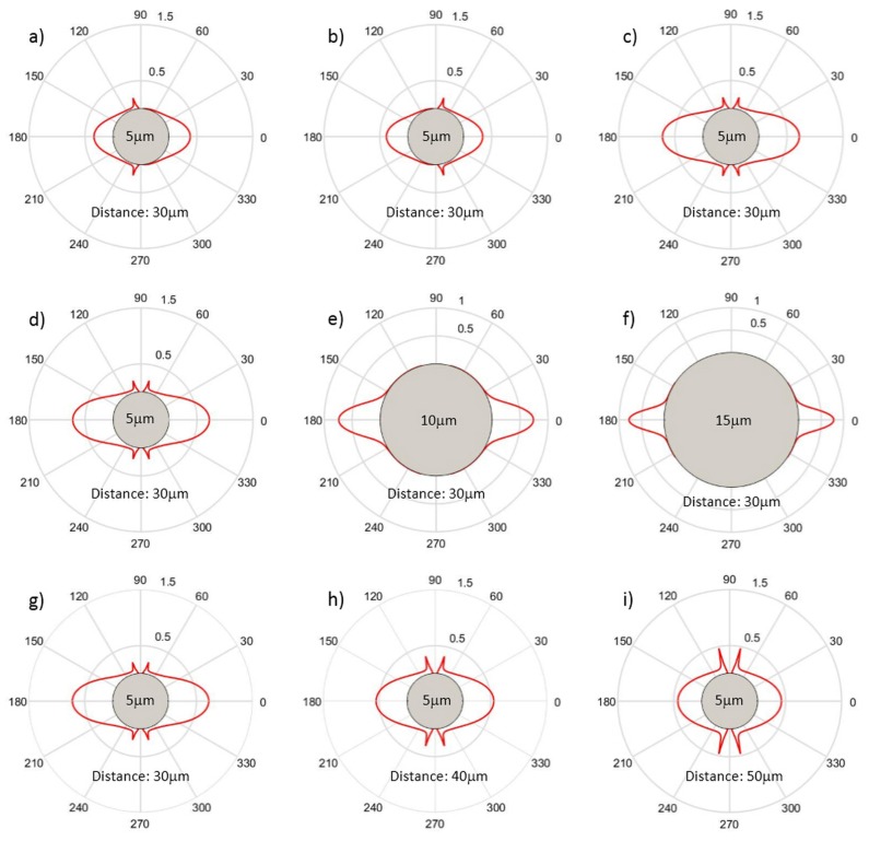
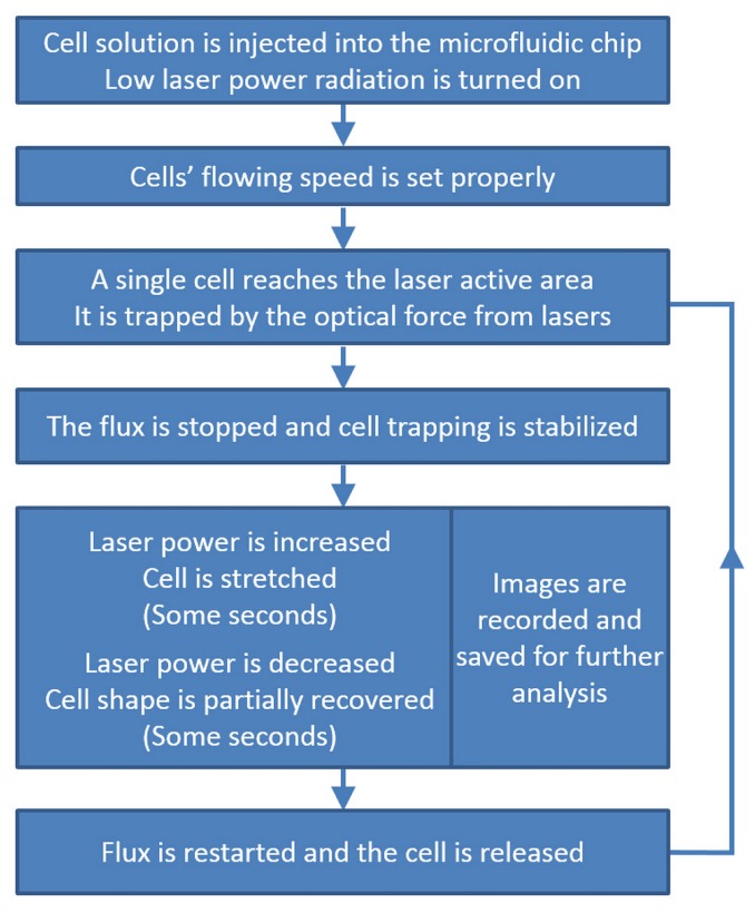
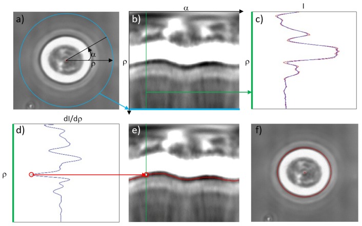
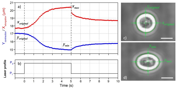
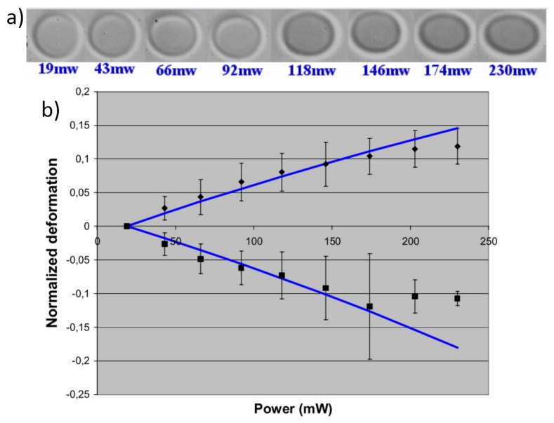
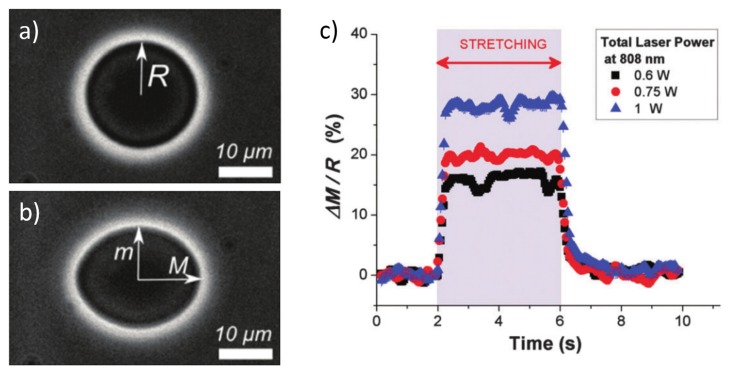
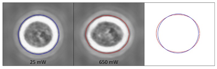
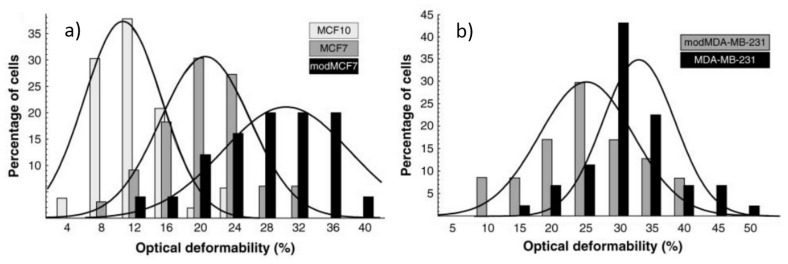
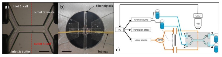
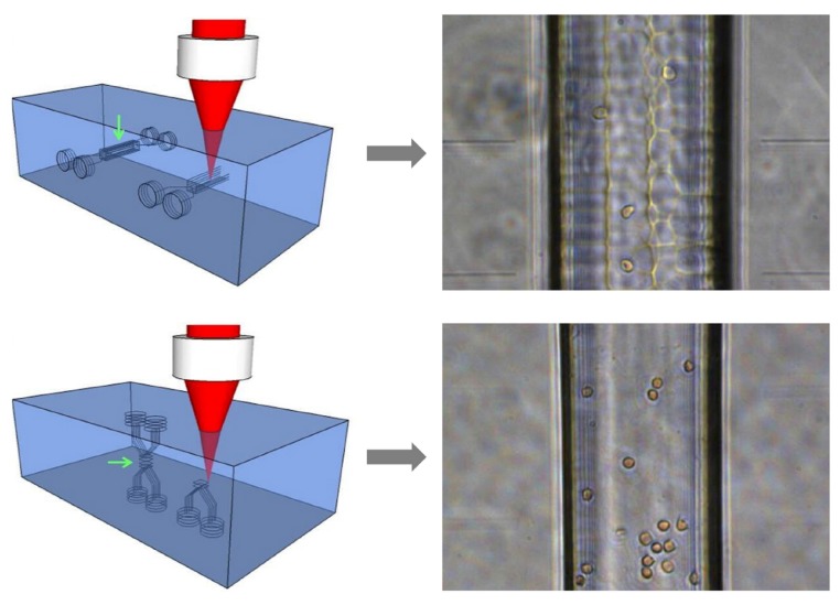
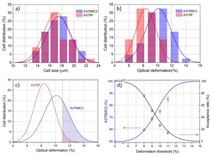
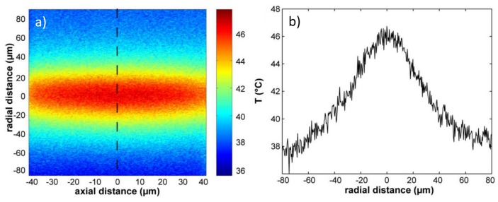
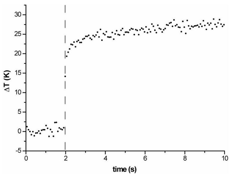
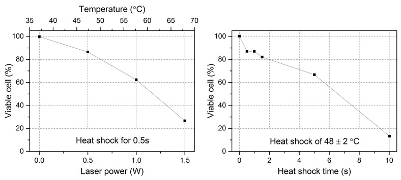
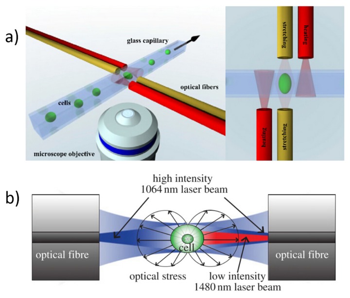
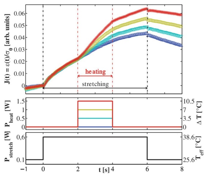
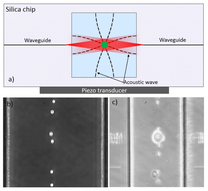
This paper presents a comprehensive review of the development of the optical stretcher, a powerful optofluidic device for single cell mechanical study by using optical force induced cell stretching. The different techniques and the different materials for the fabrication of the optical stretcher are first summarized. A short description of the optical-stretching mechanism is then given, highlighting the optical force calculation and the cell optical deformability characterization. Subsequently, the implementations of the optical stretcher in various cell-mechanics studies are shown on different types of cells. Afterwards, two new advancements on optical stretcher applications are also introduced: the active cell sorting based on cell mechanical characterization and the temperature effect on cell stretching measurement from laser-induced heating. Two examples of new functionalities developed with the optical stretcher are also included. Finally, the current major limitation and the future development possibilities are discussed.

摘要

本文全面综述了光镊的发展,光镊是一种强大的光流体装置,用于通过光力诱导细胞拉伸进行单细胞力学研究。首先总结了制造光镊的不同技术和不同材料。然后简要描述了光镊拉伸机制,重点介绍了光力计算和细胞光学变形性表征。随后,展示了光镊在各种细胞力学研究中对不同类型细胞的应用。之后,还介绍了光镊应用的两项新进展:基于细胞力学表征的主动细胞分选以及激光诱导加热对细胞拉伸测量的温度效应。文中还包括了利用光镊开发的两项新功能示例。最后,讨论了当前的主要局限性和未来的发展可能性。

https://cdn.ncbi.nlm.nih.gov/pmc/blobs/71c7/6189960/30daad48cccd/micromachines-07-00090-g025.jpg
https://cdn.ncbi.nlm.nih.gov/pmc/blobs/71c7/6189960/981eae933764/micromachines-07-00090-g001.jpg
https://cdn.ncbi.nlm.nih.gov/pmc/blobs/71c7/6189960/617afd4f9ed2/micromachines-07-00090-g002.jpg
https://cdn.ncbi.nlm.nih.gov/pmc/blobs/71c7/6189960/efb68f1280e8/micromachines-07-00090-g003.jpg
https://cdn.ncbi.nlm.nih.gov/pmc/blobs/71c7/6189960/dc7862db77db/micromachines-07-00090-g004.jpg
https://cdn.ncbi.nlm.nih.gov/pmc/blobs/71c7/6189960/aec73daa3060/micromachines-07-00090-g005.jpg
https://cdn.ncbi.nlm.nih.gov/pmc/blobs/71c7/6189960/b3c4f4f5f78c/micromachines-07-00090-g006.jpg
https://cdn.ncbi.nlm.nih.gov/pmc/blobs/71c7/6189960/d8adf00d1264/micromachines-07-00090-g007.jpg
https://cdn.ncbi.nlm.nih.gov/pmc/blobs/71c7/6189960/20c15afb3fe5/micromachines-07-00090-g008.jpg
https://cdn.ncbi.nlm.nih.gov/pmc/blobs/71c7/6189960/bcb9f5cfff26/micromachines-07-00090-g009.jpg
https://cdn.ncbi.nlm.nih.gov/pmc/blobs/71c7/6189960/45db9a66871d/micromachines-07-00090-g010.jpg
https://cdn.ncbi.nlm.nih.gov/pmc/blobs/71c7/6189960/47658c131c7c/micromachines-07-00090-g011.jpg
https://cdn.ncbi.nlm.nih.gov/pmc/blobs/71c7/6189960/5ccf83d58961/micromachines-07-00090-g012.jpg
https://cdn.ncbi.nlm.nih.gov/pmc/blobs/71c7/6189960/5ca5b660f4ce/micromachines-07-00090-g013.jpg
https://cdn.ncbi.nlm.nih.gov/pmc/blobs/71c7/6189960/455e8dd0dfa1/micromachines-07-00090-g014.jpg
https://cdn.ncbi.nlm.nih.gov/pmc/blobs/71c7/6189960/556bf34ecf77/micromachines-07-00090-g015.jpg
https://cdn.ncbi.nlm.nih.gov/pmc/blobs/71c7/6189960/50c68c250e82/micromachines-07-00090-g016.jpg
https://cdn.ncbi.nlm.nih.gov/pmc/blobs/71c7/6189960/aade12c74b05/micromachines-07-00090-g017.jpg
https://cdn.ncbi.nlm.nih.gov/pmc/blobs/71c7/6189960/2018d74bffc0/micromachines-07-00090-g018.jpg
https://cdn.ncbi.nlm.nih.gov/pmc/blobs/71c7/6189960/8d0d2593848e/micromachines-07-00090-g019.jpg
https://cdn.ncbi.nlm.nih.gov/pmc/blobs/71c7/6189960/a5616cea4692/micromachines-07-00090-g020.jpg
https://cdn.ncbi.nlm.nih.gov/pmc/blobs/71c7/6189960/a581d30cabe1/micromachines-07-00090-g021.jpg
https://cdn.ncbi.nlm.nih.gov/pmc/blobs/71c7/6189960/fca98093f6bf/micromachines-07-00090-g022.jpg
https://cdn.ncbi.nlm.nih.gov/pmc/blobs/71c7/6189960/1090cfc87bc2/micromachines-07-00090-g023.jpg
https://cdn.ncbi.nlm.nih.gov/pmc/blobs/71c7/6189960/c674258b2811/micromachines-07-00090-g024.jpg
https://cdn.ncbi.nlm.nih.gov/pmc/blobs/71c7/6189960/30daad48cccd/micromachines-07-00090-g025.jpg

相似文献

1
A Comprehensive Review of Optical Stretcher for Cell Mechanical Characterization at Single-Cell Level.单细胞水平下用于细胞力学特性表征的光镊技术综述
Micromachines (Basel). 2016 May 13;7(5):90. doi: 10.3390/mi7050090.
2
An optofluidic "tweeze-and-drag" cell stretcher in a microfluidic channel.微流控通道中的光流控“夹拉”细胞拉伸器。
Lab Chip. 2020 Feb 7;20(3):601-613. doi: 10.1039/c9lc01026b. Epub 2020 Jan 7.
3
High-throughput linear optical stretcher for mechanical characterization of blood cells.用于血细胞力学特性表征的高通量线性光镊
Cytometry A. 2016 Apr;89(4):391-7. doi: 10.1002/cyto.a.22794. Epub 2015 Nov 13.
4
The Role of the Optical Stretcher Is Crucial in the Investigation of Cell Mechanics Regulating Cell Adhesion and Motility.光镊在调节细胞黏附与运动的细胞力学研究中起着至关重要的作用。
Front Cell Dev Biol. 2019 Sep 4;7:184. doi: 10.3389/fcell.2019.00184. eCollection 2019.
5
Deformation of phospholipid vesicles in an optical stretcher.光学镊子中磷脂囊泡的变形
Soft Matter. 2015 Aug 14;11(30):6075-88. doi: 10.1039/c5sm00562k. Epub 2015 Jul 2.
6
Investigation of temperature effect on cell mechanics by optofluidic microchips.用光流控微芯片研究温度对细胞力学的影响。
Biomed Opt Express. 2015 Jul 23;6(8):2991-6. doi: 10.1364/BOE.6.002991. eCollection 2015 Aug 1.
7
Femtosecond laser fabricated monolithic chip for optical trapping and stretching of single cells.用于单细胞光镊捕获和拉伸的飞秒激光制造单片芯片。
Opt Express. 2010 Mar 1;18(5):4679-88. doi: 10.1364/OE.18.004679.
8
Optomechanical measurement of the role of lamins in whole cell deformability.核纤层蛋白在全细胞变形性中作用的光机械测量
J Biophotonics. 2017 Dec;10(12):1657-1664. doi: 10.1002/jbio.201600198. Epub 2017 May 9.
9
The optical stretcher: a novel laser tool to micromanipulate cells.光学镊子:一种用于细胞微操作的新型激光工具。
Biophys J. 2001 Aug;81(2):767-84. doi: 10.1016/S0006-3495(01)75740-2.
10
A monolithic glass chip for active single-cell sorting based on mechanical phenotyping.基于机械表型分析的活性单细胞分选用整体式玻璃芯片
Lab Chip. 2015 Mar 7;15(5):1267-75. doi: 10.1039/c4lc01196a.

引用本文的文献

1
Shape-Modified Ultrathin Glass Sheet Cantilever for Precise Single Cell Stiffness Measurement.用于精确测量单细胞刚度的形状修饰超薄玻璃板悬臂梁
ACS Meas Sci Au. 2024 Dec 4;5(3):277-286. doi: 10.1021/acsmeasuresciau.4c00074. eCollection 2025 Jun 18.
2
Effects of Shear and Extensional Stresses on Cells: Investigation in a Spiral Microchannel and Contraction-Expansion Arrays.剪切应力和拉伸应力对细胞的影响:在螺旋微通道和收缩-扩张阵列中的研究
ACS Biomater Sci Eng. 2025 Jun 9;11(6):3249-3261. doi: 10.1021/acsbiomaterials.5c00555. Epub 2025 May 28.
3
Softness or Stiffness What Contributes to Cancer and Cancer Metastasis?

本文引用的文献

1
A comprehensive strategy for the analysis of acoustic compressibility and optical deformability on single cells.一种用于分析单细胞声学压缩性和光学可变形性的综合策略。
Sci Rep. 2016 Apr 4;6:23946. doi: 10.1038/srep23946.
2
Deformability based sorting of red blood cells improves diagnostic sensitivity for malaria caused by Plasmodium falciparum.基于变形性的红细胞分选提高了对恶性疟原虫引起的疟疾的诊断敏感性。
Lab Chip. 2016 Feb 21;16(4):645-54. doi: 10.1039/c5lc01248a. Epub 2016 Jan 15.
3
Extracting Cell Stiffness from Real-Time Deformability Cytometry: Theory and Experiment.
柔软还是坚硬?是什么导致了癌症及癌症转移?
Cells. 2025 Apr 12;14(8):584. doi: 10.3390/cells14080584.
4
Integrated Microfluidics for Single-Cell Separation and On-Chip Analysis: Novel Applications and Recent Advances.用于单细胞分离和芯片分析的集成微流控技术:新应用与最新进展
Small Sci. 2024 Feb 2;4(4):2300206. doi: 10.1002/smsc.202300206. eCollection 2024 Apr.
5
Narrow-Gap Rheometry: A Novel Method for Measuring Cell Mechanics.窄缝流变学:一种测量细胞力学的新方法。
Cells. 2022 Jun 23;11(13):2010. doi: 10.3390/cells11132010.
6
Separation efficiency maximization in acoustofluidic systems: study of the sample launch-position.声流控系统中的分离效率最大化:样品注入位置研究
RSC Adv. 2018 Nov 20;8(68):38955-38964. doi: 10.1039/c8ra08860h. eCollection 2018 Nov 16.
7
On-chip integrated optical stretching and electrorotation enabling single-cell biophysical analysis.用于单细胞生物物理分析的片上集成光镊和介电电泳技术。
Microsyst Nanoeng. 2020 Jun 15;6:57. doi: 10.1038/s41378-020-0162-2. eCollection 2020.
8
Measurements of Cellular Forces and their Importance in the Lung-From the Sub- to the Multicellular Scale.细胞力的测量及其在肺中的重要性——从亚细胞尺度到多细胞尺度
Life (Basel). 2021 Jul 14;11(7):691. doi: 10.3390/life11070691.
9
Rapid computational cell-rotation around arbitrary axes in 3D with multi-core fiber.利用多核光纤在三维空间中围绕任意轴进行快速计算细胞旋转。
Biomed Opt Express. 2021 May 17;12(6):3423-3437. doi: 10.1364/BOE.423035. eCollection 2021 Jun 1.
10
The Mechanical Fingerprint of Circulating Tumor Cells (CTCs) in Breast Cancer Patients.乳腺癌患者循环肿瘤细胞(CTCs)的机械指纹图谱
Cancers (Basel). 2021 Mar 5;13(5):1119. doi: 10.3390/cancers13051119.
从实时可变形性细胞计数法中提取细胞硬度:理论与实验
Biophys J. 2015 Nov 17;109(10):2023-36. doi: 10.1016/j.bpj.2015.09.006.
4
Ezrin is a Major Regulator of Membrane Tension in Epithelial Cells.埃兹蛋白是上皮细胞膜张力的主要调节因子。
Sci Rep. 2015 Oct 5;5:14700. doi: 10.1038/srep14700.
5
Investigation of temperature effect on cell mechanics by optofluidic microchips.用光流控微芯片研究温度对细胞力学的影响。
Biomed Opt Express. 2015 Jul 23;6(8):2991-6. doi: 10.1364/BOE.6.002991. eCollection 2015 Aug 1.
6
Deformation of phospholipid vesicles in an optical stretcher.光学镊子中磷脂囊泡的变形
Soft Matter. 2015 Aug 14;11(30):6075-88. doi: 10.1039/c5sm00562k. Epub 2015 Jul 2.
7
An optofluidic constriction chip for monitoring metastatic potential and drug response of cancer cells.一种用于监测癌细胞转移潜能和药物反应的光流控收缩芯片。
Integr Biol (Camb). 2015 Apr;7(4):477-84. doi: 10.1039/c5ib00023h.
8
Real-time deformability cytometry: on-the-fly cell mechanical phenotyping.实时变形细胞术:实时细胞力学表型分析。
Nat Methods. 2015 Mar;12(3):199-202, 4 p following 202. doi: 10.1038/nmeth.3281. Epub 2015 Feb 2.
9
An integrated optofluidic device for single-cell sorting driven by mechanical properties.基于机械特性的用于单细胞分选的集成光流控装置。
Lab Chip. 2015 Mar 7;15(5):1262-6. doi: 10.1039/c4lc01496k.
10
A monolithic glass chip for active single-cell sorting based on mechanical phenotyping.基于机械表型分析的活性单细胞分选用整体式玻璃芯片
Lab Chip. 2015 Mar 7;15(5):1267-75. doi: 10.1039/c4lc01196a.