Department of Gastroenterology, The Second Affiliated Hospital of Nanchang University, Nanchang, Jiangxi, China (mainland).
Department of Electrocardiogram Diagnosis, The Second Affiliated Hospital of Nanchang University, Nanchang, Jiangxi, China (mainland).
Med Sci Monit. 2018 Nov 9;24:8015-8021. doi: 10.12659/MSM.910944.
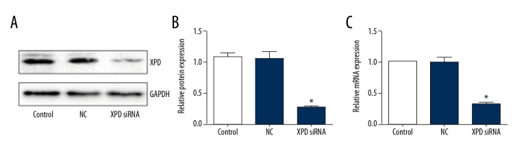
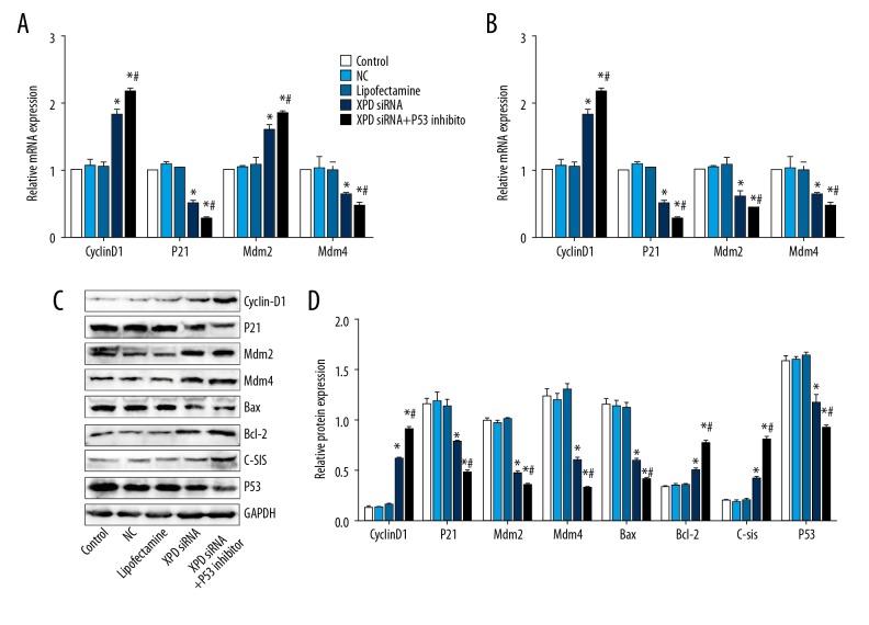
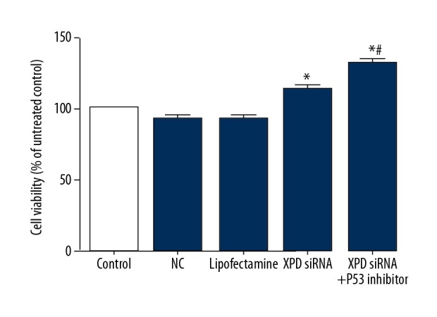
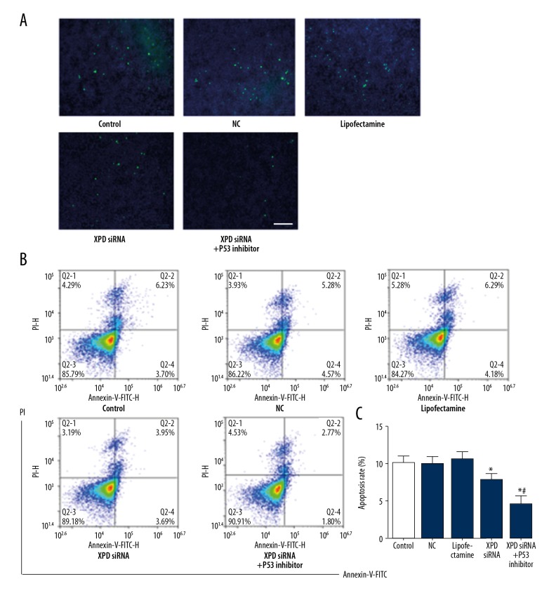
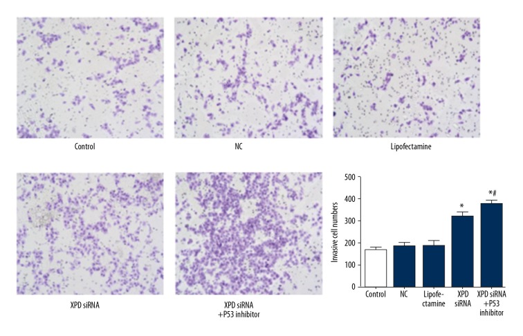
BACKGROUND This study investigated the effect of xeroderma pigmentosum group D (XPD) silencing on the growth of hepatoma cells and assessed the mechanisms. MATERIAL AND METHODS XPD gene was silenced by siRNA in hepatoma cells. The experiments were randomly divided into a control group, a liposome control group, a negative control (NC) group, an XPD siRNA group, and an XPD siRNA + P53 inhibitor group. 3-(4,5-Dimethylthiazol-2-yl)-2,5-Diphenyltetrazolium Bromide (MTT) was used to detect cell viability 24 h after gene silencing and treatments. Terminal deoxynucleotidyl transferases (TdT)-mediated dUTP nick-end labeling (TUNEL) and flow cytometry were used to detect apoptosis. Invasive ability was detected by Transwell assay. Additionally, the expression of mouse double-minute 2 homolog (Mdm2), mouse double-minute 4 homolog (Mdm4), CyclinD1, P21, Bax, P53, C-sis, and Bcl-2 was detected by real-time polymerase chain reaction and Western blotting. RESULTS Compared with the NC group, XPD siRNA significantly reduced XPD expression at both mRNA and protein levels. XPD siRNA significantly promoted cell proliferation, reduced apoptosis, and promoted cell invasive ability. Expression of CyclinD1, Bcl-2, and C-sis increased significantly after XPD silencing, while the expression of P21, Mdm2, Mdm4, Bax, and P53 significantly decreased (vs. NC, P<0.05). Importantly, P53 inhibitor (1 μM bpV) further enhanced the effect of XPD silencing (vs. XPD silencing, P<0.05). CONCLUSIONS Our data revealed that XPD silencing promoted growth of hepatoma cells by reducing P53 expression.
本研究旨在探讨 Xeroderma Pigmentosum Group D (XPD) 沉默对肝癌细胞生长的影响,并探讨其机制。
采用 siRNA 沉默肝癌细胞中的 XPD 基因。实验随机分为对照组、脂质体对照组、阴性对照组(NC 组)、XPD siRNA 组和 XPD siRNA+P53 抑制剂组。基因沉默和处理 24 小时后,采用 3-(4,5-二甲基噻唑-2-基)-2,5-二苯基四氮唑溴盐(MTT)检测细胞活力。末端脱氧核苷酸转移酶(TdT)介导的 dUTP 缺口末端标记(TUNEL)和流式细胞术检测细胞凋亡。Transwell 检测侵袭能力。实时聚合酶链反应和 Western blot 检测小鼠双微体 2 同源物(Mdm2)、小鼠双微体 4 同源物(Mdm4)、细胞周期蛋白 D1(CyclinD1)、P21、Bax、P53、C-sis 和 Bcl-2 的表达。
与 NC 组相比,XPD siRNA 显著降低了 XPD 在 mRNA 和蛋白水平的表达。XPD siRNA 显著促进细胞增殖,减少细胞凋亡,促进细胞侵袭能力。XPD 沉默后,CyclinD1、Bcl-2 和 C-sis 的表达明显增加,而 P21、Mdm2、Mdm4、Bax 和 P53 的表达明显降低(与 NC 组相比,P<0.05)。重要的是,P53 抑制剂(1 μM bpV)进一步增强了 XPD 沉默的作用(与 XPD 沉默相比,P<0.05)。
我们的数据表明,XPD 沉默通过降低 P53 表达促进肝癌细胞生长。