Department of Cardiology, Beijing Key Laboratory of Early Prediction and Intervention of Acute Myocardial Infarction, Center for Cardiovascular Translational Research, Peking University People's Hospital, Beijing 100044, China.
Chin Med J (Engl). 2018 Nov 20;131(22):2726-2733. doi: 10.4103/0366-6999.245271.
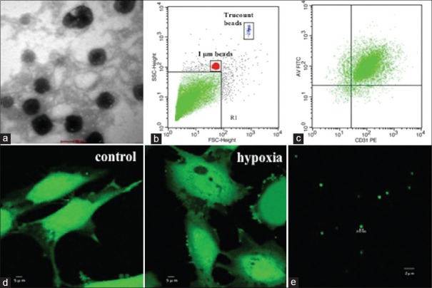
Microparticles (MPs) are small extracellular plasma membrane particles shed by activated and apoptotic cells, which are involved in the development of atherosclerosis. Our previous study found that microRNA (miR)-19b encapsulated within endothelial MPs (EMPs) may contribute to the upregulation of circulating miR-19b in unstable angina patients. Hypoxia is involved in atherosclerosis as a critical pathological stimulus. However, it still remains unclear whether the increase of miR-19b levels in EMPs is related to hypoxia and if the effect of miR-19b - wrapped within EMPs - stimulates hypoxia on vascular endothelial cells. This study aimed to explore the changes of miR-19b in EMPs induced by hypoxia as well as their effects on endothelial cells.
Human umbilical vein endothelial cells (HUVECs) were cultured in vitro and arranged to harvest EMPs in two parts: the first part consisted of EMP and EMP and the second part included EMP, EMP, and EMP. Cell migration was detected by scratch migration and transwell chamber migration. Angiogenesis was assessed by tube formation assays. Furthermore, we predicted the target gene of miR-19b by bioinformatics analysis, and luciferase assay was used to verify the targeted gene of miR-19b. Data were analyzed by one-way analysis of variance. Student's t-test was used when two groups were compared.
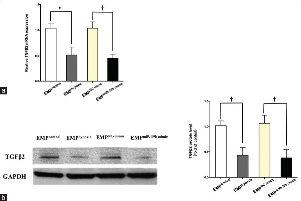
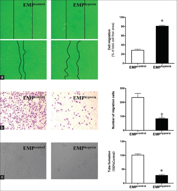
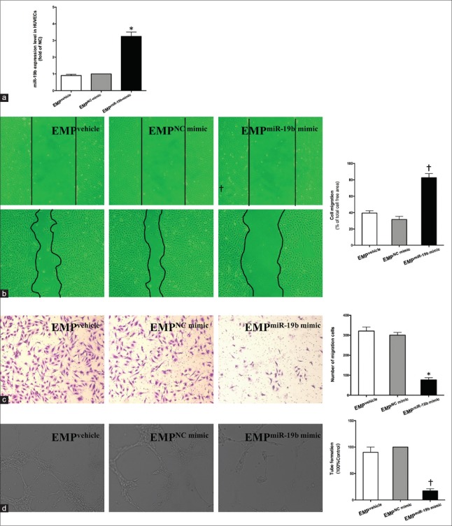
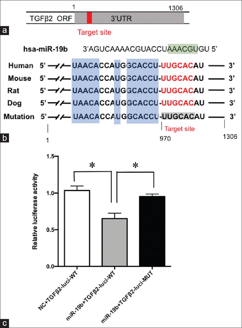
Compared with EMP- and EMP-inhibited migration of cells by scratch migration assay (80.77 ± 1.10 vs. 28.37 ± 1.40, P < 0. 001) and transwell chamber migration assay (83.00 ± 3.46 vs. 235.00 ± 16.52, P < 0.01), the number of tube formations was markedly reduced by 70% in the EMP group (P < 0.001) in vitro analysis of HUVECs. Meanwhile, a strong inhibition of migration and tube formation of HUVECs in the presence of miR-19b-enriched EMP was observed. This effect might be due to the delivery of miR-19b in EMPs. Transforming growth factor-β2 (TGFβ2) was predicted to be one of the target genes of miR-19b, and we further confirmed that TGFβ2 was a direct target gene of miR-19b using the luciferase assay. The expression of TGFβ2 in HUVECs was inhibited by treatment with EMP and EMP.
MiR-19b in EMPs induced by hypoxia could reduce endothelial cell migration and angiogenesis by downregulating TGFβ2 expression, which may have inhibited the progression of atherosclerosis.
微粒(MPs)是由激活和凋亡细胞释放的小细胞外质膜颗粒,参与动脉粥样硬化的发展。我们之前的研究发现,内皮 MPs(EMPs)中包裹的 microRNA(miR)-19b 可能导致不稳定型心绞痛患者循环 miR-19b 水平升高。缺氧作为一种关键的病理刺激参与动脉粥样硬化。然而,目前尚不清楚 EMPs 中 miR-19b 水平的增加是否与缺氧有关,以及包裹在 EMPs 中的 miR-19b 的作用是否会刺激血管内皮细胞缺氧。本研究旨在探讨缺氧诱导的 EMPs 中 miR-19b 的变化及其对内皮细胞的影响。
体外培养人脐静脉内皮细胞(HUVECs),分为两部分收集 EMPs:第一部分包括 EMP 和 EMP,第二部分包括 EMP、EMP 和 EMP。划痕迁移和 Transwell 室迁移检测细胞迁移。管形成试验评估血管生成。此外,我们通过生物信息学分析预测了 miR-19b 的靶基因,并通过荧光素酶报告基因实验验证了 miR-19b 的靶基因。采用单因素方差分析进行数据分析。当比较两组时,使用 Student's t-test。
与 EMP 和 EMP 抑制划痕迁移试验(80.77 ± 1.10 对 28.37 ± 1.40,P < 0.001)和 Transwell 室迁移试验(83.00 ± 3.46 对 235.00 ± 16.52,P < 0.01)的细胞迁移相比, EMP 组(P < 0.001)体外分析的 HUVECs 中管形成数量明显减少。同时,在 miR-19b 富集的 EMP 存在的情况下,HUVECs 的迁移和管形成受到强烈抑制。这种作用可能是由于 EMP 中 miR-19b 的传递。转化生长因子-β2(TGFβ2)被预测为 miR-19b 的靶基因之一,我们进一步通过荧光素酶报告基因实验证实了 TGFβ2 是 miR-19b 的直接靶基因。EMP 和 EMP 处理抑制了 HUVECs 中 TGFβ2 的表达。
缺氧诱导的 EMPs 中的 miR-19b 通过下调 TGFβ2 的表达减少内皮细胞迁移和血管生成,可能抑制了动脉粥样硬化的进展。