• 文献检索
  • 文档翻译
  • 深度研究
  • 学术资讯
  • Suppr Zotero 插件Zotero 插件
  • 邀请有礼
  • 套餐&价格
  • 历史记录
应用&插件
Suppr Zotero 插件Zotero 插件浏览器插件Mac 客户端Windows 客户端微信小程序
定价
高级版会员购买积分包购买API积分包
服务
文献检索文档翻译深度研究API 文档MCP 服务
关于我们
关于 Suppr公司介绍联系我们用户协议隐私条款
关注我们

Suppr 超能文献

核心技术专利:CN118964589B侵权必究
粤ICP备2023148730 号-1Suppr @ 2026

文献检索

告别复杂PubMed语法,用中文像聊天一样搜索,搜遍4000万医学文献。AI智能推荐,让科研检索更轻松。

立即免费搜索

文件翻译

保留排版,准确专业,支持PDF/Word/PPT等文件格式,支持 12+语言互译。

免费翻译文档

深度研究

AI帮你快速写综述,25分钟生成高质量综述,智能提取关键信息,辅助科研写作。

立即免费体验

直肠癌弥散加权成像的可重复性和癌症特征:b 值分布研究的影响。

Diffusion-weighted imaging of rectal cancer on repeatability and cancer characterization: an effect of b-value distribution study.

机构信息

Department of Radiology, Changhai Hospital of Shanghai, The Second Military Medical University, No.168 Changhai Road, Shanghai, 200433, China.

出版信息

Cancer Imaging. 2018 Nov 15;18(1):43. doi: 10.1186/s40644-018-0177-1.

DOI:10.1186/s40644-018-0177-1
PMID:30442202
原文链接:https://pmc.ncbi.nlm.nih.gov/articles/PMC6238345/
Abstract

BACKGROUND

To explore the effect of b-value distributions on the repeatability and diagnostic performance of the ADC value in rectal cancer patients using multiple b-values and mono-exponential model diffusion-weighted imaging (DWI).

METHODS

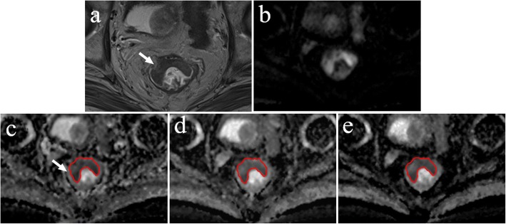
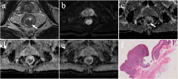
Thirty-two preoperative rectal cancer patients, without receiving neoadjuvant therapy, were scanned on a 3 Tesla magnetic resonance imaging scanner using DWI with 10 b-values ranging from 0 to 2000 s/mm. The apparent diffusion coefficient (ADC) value was calculated using a mono-exponential model and 31 b-value combinations consisting of 2 to 10 b-values were explored. Regions of interest with the maximum cross-sectional tumour size were outlined on the ADC map by two independent observers. Intraclass correlation coefficients (ICC), coefficient of variation (CV), and Bland-Altman plots between the two observers were calculated and evaluated to determine repeatability. Areas under receiver operating characteristic curves (AUCs) were evaluated for rectal cancer characterization. Correlations between the mean ADC values and T stage were assessed using the Spearman correlation coefficient (ρ). α (= ICC + AUC + |ρ|- CV - |bias|) was defined and used to assess the optimal b-value distribution.

RESULTS

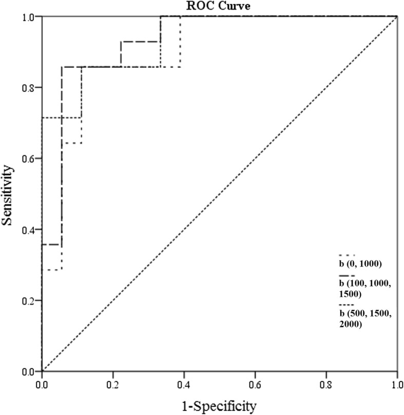
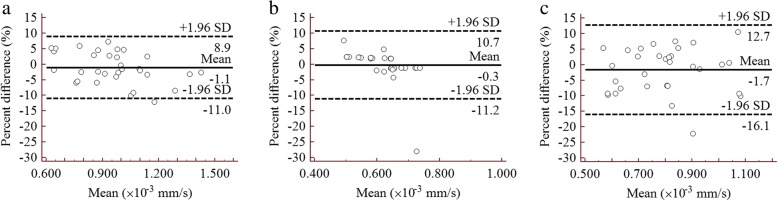
Postoperative pathology tests revealed 4 patients with T1, 10 patients with T2, and 18 patients with T3 stages. There were no significant difference in age and sex between the two groups (T1-2 vs. T3). Excellent reproducibility was observed for ADC values between two observers with ICC and CV values ranging from 0.920 to 0.998, and 1.475 to 5.568%, respectively. The mean percent difference and ρ between the paired measurements was ranged from - 2.7 to 1.2% and from - 0.759 to - 0.407, respectively. The b-value combinations with the top three α values were b(0, 1000 s/mm), b(500, 1500, 2000 s/mm) and b(100, 1000, 1500 s/mm) for α = 2.581, 2.571 and 2.569, respectively.

CONCLUSIONS

The number of b-values and their distributions influenced the repeatability of the ADC values and their diagnostic performance. The optimal b-value combination was 0 and 1000 s/mm for DWI examination of rectal cancer patients.

摘要

背景

本研究旨在探讨使用多 b 值和单指数模型扩散加权成像(DWI)时,b 值分布对直肠癌患者 ADC 值重复性和诊断性能的影响。

方法

32 例术前直肠癌患者未接受新辅助治疗,在 3.0T 磁共振成像扫描仪上进行 DWI 扫描,b 值范围为 0 至 2000 s/mm,共 10 个。使用单指数模型计算表观扩散系数(ADC)值,并探讨了由 2 至 10 个 b 值组成的 31 种 b 值组合。两位独立观察者在 ADC 图上勾画最大肿瘤横截面积的感兴趣区。计算两位观察者之间的组内相关系数(ICC)、变异系数(CV)和 Bland-Altman 图,并进行评估,以确定重复性。评估受试者工作特征曲线(ROC)下面积(AUC)以用于直肠癌特征描述。使用 Spearman 相关系数(ρ)评估平均 ADC 值与 T 分期之间的相关性。定义了α(= ICC+AUC+|ρ|-CV-|偏倚|)并用于评估最佳 b 值分布。

结果

术后病理检查显示 4 例患者为 T1 期,10 例患者为 T2 期,18 例患者为 T3 期。两组间(T1-2 与 T3)在年龄和性别上无显著差异。两位观察者之间的 ADC 值具有极好的可重复性,ICC 值范围为 0.920 至 0.998,CV 值范围为 1.475 至 5.568%。配对测量的平均百分比差异和ρ值范围分别为-2.7%至 1.2%和-0.759 至-0.407。α值为 2.581、2.571 和 2.569 时,最佳 b 值组合分别为 b(0、1000 s/mm)、b(500、1500、2000 s/mm)和 b(100、1000、1500 s/mm)。

结论

b 值的数量及其分布影响 ADC 值的重复性及其诊断性能。对于直肠癌患者的 DWI 检查,最佳 b 值组合为 0 和 1000 s/mm。

https://cdn.ncbi.nlm.nih.gov/pmc/blobs/740d/6238345/a8ef8d7cd373/40644_2018_177_Fig6_HTML.jpg
https://cdn.ncbi.nlm.nih.gov/pmc/blobs/740d/6238345/8e0c7b28dc10/40644_2018_177_Fig1_HTML.jpg
https://cdn.ncbi.nlm.nih.gov/pmc/blobs/740d/6238345/f9a01996f936/40644_2018_177_Fig2_HTML.jpg
https://cdn.ncbi.nlm.nih.gov/pmc/blobs/740d/6238345/e30c637713a4/40644_2018_177_Fig3_HTML.jpg
https://cdn.ncbi.nlm.nih.gov/pmc/blobs/740d/6238345/815e43fbeb19/40644_2018_177_Fig4_HTML.jpg
https://cdn.ncbi.nlm.nih.gov/pmc/blobs/740d/6238345/8b96d52c1e4c/40644_2018_177_Fig5_HTML.jpg
https://cdn.ncbi.nlm.nih.gov/pmc/blobs/740d/6238345/a8ef8d7cd373/40644_2018_177_Fig6_HTML.jpg

相似文献

1
Diffusion-weighted imaging of rectal cancer on repeatability and cancer characterization: an effect of b-value distribution study.直肠癌弥散加权成像的可重复性和癌症特征:b 值分布研究的影响。
Cancer Imaging. 2018 Nov 15;18(1):43. doi: 10.1186/s40644-018-0177-1.
2
The usefulness of b value threshold map in the evaluation of rectal adenocarcinoma.b 值阈值图在直肠腺癌评估中的应用价值。
Abdom Radiol (NY). 2020 Feb;45(2):332-341. doi: 10.1007/s00261-019-02272-0.
3
Prediction of different stages of rectal cancer: Texture analysis based on diffusion-weighted images and apparent diffusion coefficient maps.预测直肠癌不同阶段:基于扩散加权图像和表观扩散系数图的纹理分析。
World J Gastroenterol. 2020 May 7;26(17):2082-2096. doi: 10.3748/wjg.v26.i17.2082.
4
Diffusion-weighted imaging of prostate cancer: effect of b-value distribution on repeatability and cancer characterization.前列腺癌的扩散加权成像:b值分布对重复性和癌症特征描述的影响。
Magn Reson Imaging. 2015 Dec;33(10):1212-1218. doi: 10.1016/j.mri.2015.07.004. Epub 2015 Jul 26.
5
Repeatability of diffusion-weighted imaging in rectal cancer.直肠癌扩散加权成像的可重复性
J Magn Reson Imaging. 2014 Jul;40(1):146-50. doi: 10.1002/jmri.24337. Epub 2013 Oct 11.
6
Diffusion-weighted imaging: Apparent diffusion coefficient histogram analysis for detecting pathologic complete response to chemoradiotherapy in locally advanced rectal cancer.扩散加权成像:表观扩散系数直方图分析用于检测局部晚期直肠癌对放化疗的病理完全缓解
J Magn Reson Imaging. 2016 Jul;44(1):212-20. doi: 10.1002/jmri.25117. Epub 2015 Dec 15.
7
Assessment and quantification of sources of variability in breast apparent diffusion coefficient (ADC) measurements at diffusion weighted imaging.扩散加权成像中乳腺表观扩散系数(ADC)测量变异性来源的评估与量化
Eur J Radiol. 2015 Sep;84(9):1729-36. doi: 10.1016/j.ejrad.2015.05.032. Epub 2015 Jun 1.
8
[Measurement reproducibility of parameters derived from introvoxel incoherent motion diffusion-weighted MRI imaging of rectal cancer].[直肠癌体素内不相干运动扩散加权磁共振成像参数的测量可重复性]
Zhonghua Zhong Liu Za Zhi. 2017 Dec 23;39(12):910-915. doi: 10.3760/cma.j.issn.0253-3766.2017.12.006.
9
Comparison of reduced field-of-view diffusion-weighted imaging (DWI) and conventional DWI techniques in the assessment of rectal carcinoma at 3.0T: Image quality and histological T staging.3.0T 下缩小视野弥散加权成像(DWI)与常规 DWI 技术在直肠癌评估中的比较:图像质量与组织学 T 分期。
J Magn Reson Imaging. 2018 Apr;47(4):967-975. doi: 10.1002/jmri.25814. Epub 2017 Jul 10.
10
Value of diffusion-weighted MRI and apparent diffusion coefficient measurements for predicting the response of locally advanced rectal cancer to neoadjuvant chemoradiotherapy.弥散加权 MRI 及其表观弥散系数测量在预测局部进展期直肠癌新辅助放化疗疗效中的价值。
Abdom Radiol (NY). 2016 Oct;41(10):1906-17. doi: 10.1007/s00261-016-0805-9.

引用本文的文献

1
Comparison of apparent diffusion coefficients of resectable mid‑high rectal adenocarcinoma and distal paracancerous tissue.可切除的中高位直肠腺癌与癌旁远端组织的表观扩散系数比较。
Oncol Lett. 2024 Dec 10;29(2):97. doi: 10.3892/ol.2024.14843. eCollection 2025 Feb.
2
Ultra-high b-value DWI in rectal cancer: image quality assessment and regional lymph node prediction based on radiomics.直肠癌中的超高b值扩散加权成像:基于影像组学的图像质量评估和区域淋巴结预测
Eur Radiol. 2025 Jan;35(1):49-60. doi: 10.1007/s00330-024-10958-3. Epub 2024 Jul 12.
3
The Multipurpose Usage of Diffusion-Weighted MRI in Rectal Cancer.

本文引用的文献

1
Image Quality Assessment of 2D 3D T2WI and Evaluation of Ultra-high b-Value (b=2,000 mm/s) DWI for Response Assessment in Rectal Cancer.二维及三维T2WI图像质量评估与超高b值(b = 2000mm²/s)弥散加权成像在直肠癌疗效评估中的应用
Anticancer Res. 2018 Feb;38(2):969-978. doi: 10.21873/anticanres.12311.
2
Quantitative Aspects of Diffusion-weighted Magnetic Resonance Imaging in Rectal Cancer Response to Neoadjuvant Therapy.直肠癌新辅助治疗反应的扩散加权磁共振成像的定量分析
Radiol Oncol. 2017 Jun 25;51(3):270-276. doi: 10.1515/raon-2017-0025. eCollection 2017 Sep.
3
Study protocol: multi-parametric magnetic resonance imaging for therapeutic response prediction in rectal cancer.
磁共振弥散加权成像在直肠癌中的多方面应用。
Medicina (Kaunas). 2023 Dec 13;59(12):2162. doi: 10.3390/medicina59122162.
4
MRI Evaluation of Rectal Cancer Lymph Node Staging Using Apparent Diffusion Coefficient.利用表观扩散系数对直肠癌淋巴结分期进行磁共振成像评估
Cureus. 2023 Sep 10;15(9):e45002. doi: 10.7759/cureus.45002. eCollection 2023 Sep.
5
Comparison of Computed and Acquired DWI in the Assessment of Rectal Cancer: Image Quality and Preoperative Staging.计算机断层扫描与采集的扩散加权成像在直肠癌评估中的比较:图像质量与术前分期
Front Oncol. 2022 Mar 18;12:788731. doi: 10.3389/fonc.2022.788731. eCollection 2022.
6
Integrated slice-specific dynamic shimming diffusion weighted imaging (DWI) for rectal Cancer detection and characterization.直肠癌检测和特征分析的集成切片特异性动态匀场弥散加权成像(DWI)
Cancer Imaging. 2021 Apr 7;21(1):32. doi: 10.1186/s40644-021-00403-9.
7
Endorectal Ultrasound and Magnetic Resonance Imaging for Rectal Cancer Staging: A Modern Multimodality Approach.用于直肠癌分期的直肠内超声和磁共振成像:一种现代多模态方法。
J Clin Med. 2021 Feb 8;10(4):641. doi: 10.3390/jcm10040641.
8
Manual and semi-automated delineation of locally advanced rectal cancer subvolumes with diffusion-weighted MRI.基于弥散加权 MRI 的局部进展期直肠癌亚体积手动和半自动勾画。
Br J Radiol. 2020 Oct 1;93(1114):20200543. doi: 10.1259/bjr.20200543. Epub 2020 Sep 2.
9
Optimized Parameters of Diffusion-Weighted MRI for Prediction of the Response to Neoadjuvant Chemoradiotherapy for Locally Advanced Rectal Cancer.扩散加权 MRI 参数优化预测局部进展期直肠癌新辅助放化疗疗效的研究
Biomed Res Int. 2019 Oct 13;2019:9392747. doi: 10.1155/2019/9392747. eCollection 2019.
10
Apparent diffusion coefficient-based histogram analysis differentiates histological subtypes of periampullary adenocarcinoma.基于表观扩散系数的直方图分析可区分壶腹周围腺癌的组织学亚型。
World J Gastroenterol. 2019 Oct 28;25(40):6116-6128. doi: 10.3748/wjg.v25.i40.6116.
研究方案:用于直肠癌治疗反应预测的多参数磁共振成像
BMC Cancer. 2017 Jul 4;17(1):465. doi: 10.1186/s12885-017-3449-4.
4
Colon Cancer, Version 1.2017, NCCN Clinical Practice Guidelines in Oncology.《结肠癌临床实践指南(2017 年版)》,NCCN 肿瘤学临床实践指南。
J Natl Compr Canc Netw. 2017 Mar;15(3):370-398. doi: 10.6004/jnccn.2017.0036.
5
Functional MRI for quantitative treatment response prediction in locally advanced rectal cancer.功能磁共振成像用于局部晚期直肠癌定量治疗反应预测
Br J Radiol. 2017 Apr;90(1072):20151078. doi: 10.1259/bjr.20151078. Epub 2017 Mar 7.
6
Comparison of non-Gaussian and Gaussian diffusion models of diffusion weighted imaging of rectal cancer at 3.0 T MRI.直肠磁共振 3.0T 成像弥散加权成像中基于非高斯和高斯扩散模型的对比研究。
Sci Rep. 2016 Dec 9;6:38782. doi: 10.1038/srep38782.
7
Diffusion-weighted imaging outside the brain: Consensus statement from an ISMRM-sponsored workshop.脑外扩散加权成像:ISMRM 主办研讨会的共识声明
J Magn Reson Imaging. 2016 Sep;44(3):521-40. doi: 10.1002/jmri.25196. Epub 2016 Feb 19.
8
Cancer statistics in China, 2015.《中国癌症统计数据 2015》
CA Cancer J Clin. 2016 Mar-Apr;66(2):115-32. doi: 10.3322/caac.21338. Epub 2016 Jan 25.
9
Apparent diffusion coefficient (ADC) measurement in ovarian tumor: Effect of region-of-interest methods on ADC values and diagnostic ability.卵巢肿瘤的表观扩散系数(ADC)测量:感兴趣区方法对ADC值及诊断能力的影响
J Magn Reson Imaging. 2016 Mar;43(3):720-5. doi: 10.1002/jmri.25011. Epub 2015 Jul 22.
10
Apparent diffusion coefficient (ADC) measurements in pancreatic adenocarcinoma: A preliminary study of the effect of region of interest on ADC values and interobserver variability.胰腺腺癌的表观扩散系数(ADC)测量:关于感兴趣区域对ADC值及观察者间变异性影响的初步研究
J Magn Reson Imaging. 2016 Feb;43(2):407-13. doi: 10.1002/jmri.25007. Epub 2015 Jul 16.