Fulong Xiao, Chao Lu, Dianjiang Zhao, Qihong Zou, Wei Zhang, Jun Zhang, Fang Han
Department of Respiratory and Critical Care Medicine, Sleep Medicine Center, Peking University People's Hospital, Beijing, China.
Department of Radiology, Peking University International Hospital, Beijing, China.
Front Neurol. 2018 Nov 2;9:936. doi: 10.3389/fneur.2018.00936. eCollection 2018.
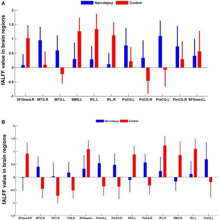
To identify narcolepsy related regional brain activity alterations compared with matched healthy controls. To determine whether these changes can be used to distinguish narcolepsy from healthy controls by recursive partitioning analysis (RPA) and receiver operating characteristic (ROC) curve analysis. Fifty-one narcolepsy with cataplexy patients (26 adults and 25 juveniles) and sixty matched heathy controls (30 adults and 30 juveniles) were recruited. All subjects underwent a resting-state functional magnetic resonance imaging scan. Fractional low-frequency fluctuations (fALFF) was used to investigate narcolepsy induced regional brain activity alterations among adult and juveniles, respectively. Recursive partitioning analysis and Receiver operating curve analysis was used to seek the ability of fALFF values within brain regions in distinguishing narcolepsy from healthy controls. Compared with healthy controls, both adult and juvenile narcolepsy had lower fALFF values in bilateral medial superior frontal gyrus, bilateral inferior parietal lobule and supra-marginal gyrus. Compared with healthy controls, both adult and juvenile narcolepsy had higher fALFF values in bilateral sensorimotor cortex and middle temporal gyrus. Also juvenile narcolepsy had higher fALFF in right putamen and right thalamus compared with healthy controls. Based on RPA and ROC curve analysis, in adult participants, fALFF differences in right medial superior frontal gyrus can discriminate narcolepsy from healthy controls with high degree of sensitivity (100%) and specificity (88.9%). In juvenile participants, fALFF differences in left superior frontal gyrus can discriminate narcolepsy from healthy controls with moderate degree of sensitivity (57.1%) and specificity (88.9%). Compared with healthy controls, both the adult and juvenile narcolepsy showed overlap brain regions in fALFF differences after case-control comparison. Furthermore, we propose that fALFF value can be a helpful imaging biomarker in distinguishing narcolepsy from healthy controls among both adults and juveniles.
为了识别与发作性睡病相关的脑区活动变化,并与匹配的健康对照进行比较。通过递归划分分析(RPA)和受试者工作特征(ROC)曲线分析,确定这些变化是否可用于区分发作性睡病与健康对照。招募了51例伴猝倒的发作性睡病患者(26例成人和25例青少年)和60例匹配的健康对照(30例成人和30例青少年)。所有受试者均接受静息态功能磁共振成像扫描。分别采用低频振幅分数(fALFF)来研究发作性睡病在成人和青少年中诱发的脑区活动变化。采用递归划分分析和受试者工作曲线分析,以寻找脑区内fALFF值区分发作性睡病与健康对照的能力。与健康对照相比,成人和青少年发作性睡病患者双侧额上回内侧、双侧顶下小叶和缘上回的fALFF值均较低。与健康对照相比,成人和青少年发作性睡病患者双侧感觉运动皮层和颞中回的fALFF值均较高。此外,与健康对照相比,青少年发作性睡病患者右侧壳核和右侧丘脑的fALFF值较高。基于RPA和ROC曲线分析,在成年参与者中,右侧额上回内侧的fALFF差异能够以高灵敏度(100%)和特异性(88.9%)区分发作性睡病与健康对照。在青少年参与者中,左侧额上回的fALFF差异能够以中等灵敏度(57.1%)和特异性(88.9%)区分发作性睡病与健康对照。与健康对照相比,成人和青少年发作性睡病在病例对照比较后,fALFF差异显示出重叠的脑区。此外,我们提出fALFF值可能是区分成人和青少年发作性睡病与健康对照的一种有用的影像学生物标志物。