Piasek Ewa, Sojka Michał, Kuczyńska Maryla, Światłowski Łukasz, Drelich-Zbroja Anna, Furmaga Olga, Jargiełło Tomasz
Student Study Group, Department of Interventional Radiology and Neuroradiology, Medical University of Lublin, Lublin, Poland.
Department of Interventional Radiology and Neuroradiology, Medical University of Lublin, Lublin, Poland.
J Ultrason. 2018;18(73):148-151. doi: 10.15557/JoU.2018.0021.
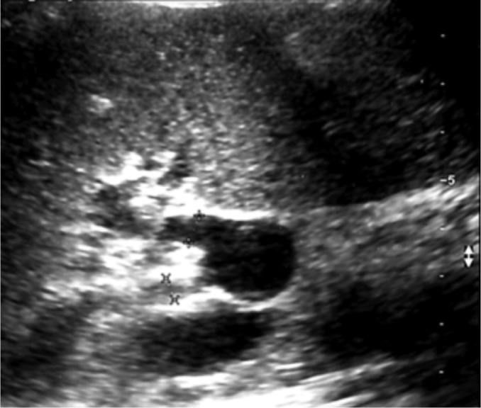
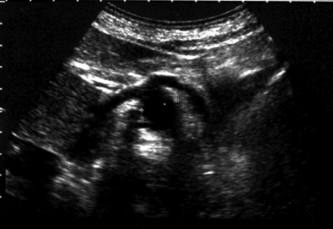
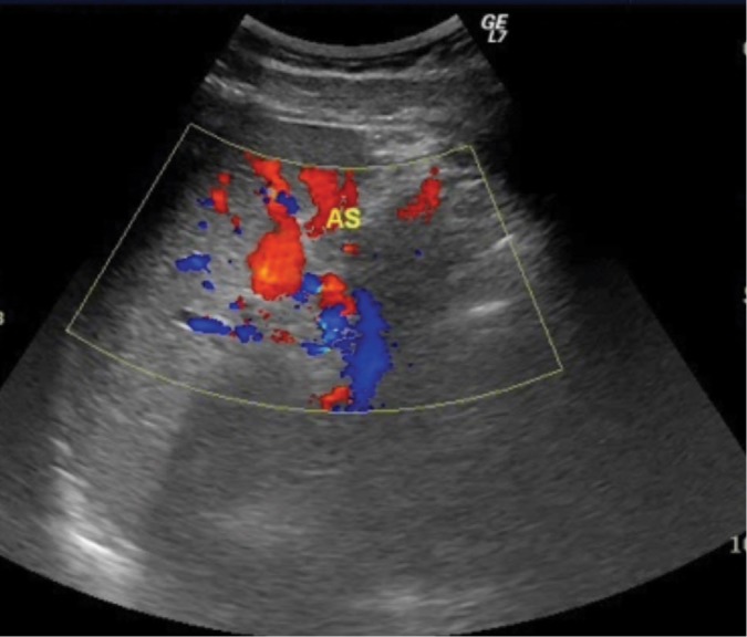
Although visceral artery aneurysms are rare, mortality due to their rupture is high, estimated at even 25-75%. That is why it is significant to detect each such lesion. Visceral artery aneurysms are usually asymptomatic and found incidentally during examinations performed for other indications. Autopsy results suggest that most asymptomatic aneurysms remain undiagnosed during lifetime. Their prevalence in the population is therefore higher. The manifestation of a ruptured aneurysm depends on its location and may involve intraperitoneal hemorrhage, gastrointestinal and portal system bleeding with concomitant portal hypertension and bleeding from esophageal varices. Wide access to diagnostic tests, for example ultrasound, computed tomography or magnetic resonance imaging, helps establish the correct diagnosis and a therapeutic plan as well as select appropriate treatment. After a procedure, the same diagnostic tools enable assessment of treatment efficacy, or are used for the monitoring of aneurysm size and detection of potential complications in cases that are ineligible for treatment. The type of treatment depends on the size of an aneurysm, the course of the disease, risk of rupture and risk associated with surgery or endovascular procedure. Endovascular treatment is preferred in most cases. Aneurysms are excluded from the circulation using embolization coils, ethylene vinyl alcohol, stents, multilayer stents, stent grafts and histoacryl glue (or a combination of these methods).
虽然内脏动脉瘤很少见,但其破裂导致的死亡率很高,估计甚至可达25% - 75%。这就是检测每一例此类病变为何重要的原因。内脏动脉瘤通常无症状,在因其他指征进行检查时偶然发现。尸检结果表明,大多数无症状动脉瘤在生前仍未被诊断出来。因此它们在人群中的患病率更高。动脉瘤破裂的表现取决于其位置,可能包括腹腔内出血、胃肠道和门静脉系统出血,伴有门静脉高压以及食管静脉曲张出血。广泛应用诊断检查,例如超声、计算机断层扫描或磁共振成像,有助于确立正确诊断和治疗方案,以及选择合适的治疗方法。手术后,同样的诊断工具可用于评估治疗效果,或在不适合治疗的病例中用于监测动脉瘤大小和检测潜在并发症。治疗类型取决于动脉瘤的大小、病程、破裂风险以及与手术或血管内介入相关的风险。大多数情况下首选血管内治疗。使用栓塞线圈、乙烯醇、支架、多层支架、覆膜支架和组织黏合剂(或这些方法的组合)将动脉瘤排除在循环之外。