Center for Life Science Research, University of Yamanashi, Chuo, Yamanashi, Japan.
Interdisciplinary Graduate School, University of Yamanashi, Kofu, Yamanashi, Japan.
Anat Sci Educ. 2019 Sep;12(5):561-571. doi: 10.1002/ase.1822. Epub 2018 Nov 19.
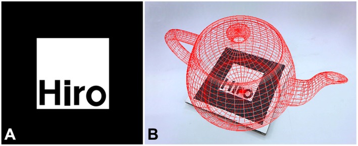
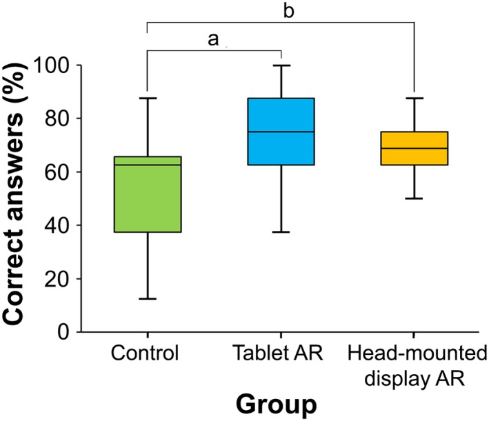
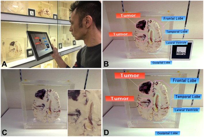
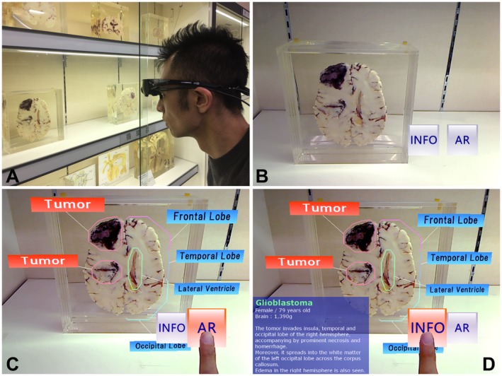
Human anatomical specimen museums are commonly used by medical, nursing, and paramedical students. Through dissection and prosection, the specimens housed in these museums allow students to appreciate the complex relationships of organs and structures in more detail than textbooks could provide. However, it may be difficult for students, particularly novices, to identify the various parts of these anatomical structures without additional explanations from a docent or supplemental illustrations. Recently, augmented reality (AR) has been used in many museum exhibits to display virtual objects in videos captured from the real world. This technology can significantly enhance the learning experience. In this study, three AR-based support systems for tours in medical specimen museums were developed, and their usability and effectiveness for learning were examined. The first system was constructed using an AR marker. This system could display virtual label information for specimens by capturing AR markers using a tablet camera. Individual AR markers were required for all specimens, but their presence in and on the prosected specimens could also be obtrusive. The second system was developed to set the specimen image itself as an image marker, as most specimens were displayed in cross section. Visitors could then obtain the label information presented by AR without any markers intruding on the display or anatomical specimens. The third system was comprised of a head-mounted display combined with a natural click interface. The system could provide visitors with an environment for the natural manipulation of virtual objects with future scalability.
人体解剖标本博物馆通常被医学、护理和辅助医疗专业的学生使用。通过解剖和切片,这些博物馆中存放的标本使学生能够更详细地了解器官和结构之间的复杂关系,这比教科书所能提供的内容更加详细。然而,对于学生,特别是初学者来说,如果没有讲解员的额外解释或补充插图,他们可能很难识别这些解剖结构的各个部分。最近,增强现实(AR)技术已在许多博物馆展览中使用,以在从现实世界中捕获的视频中显示虚拟对象。这项技术可以显著增强学习体验。在这项研究中,开发了三种基于 AR 的医学标本博物馆参观支持系统,并对它们的学习可用性和有效性进行了检验。第一个系统是使用 AR 标记构建的。该系统可以通过使用平板电脑相机捕获 AR 标记来显示标本的虚拟标签信息。每个标本都需要单独的 AR 标记,但它们在被切开的标本上和内部的存在可能会有些碍眼。第二个系统是为将标本图像本身设置为图像标记而开发的,因为大多数标本都是以横切面的形式展示的。然后,参观者可以在不干扰显示或解剖标本的情况下通过 AR 获取标签信息。第三个系统由一个头戴式显示器和一个自然点击界面组成。该系统可以为参观者提供一个环境,让他们可以对虚拟对象进行自然操作,并具有未来的可扩展性。