Davis Ryan M, Campbell Jos L, Burkitt Sean, Qiu Zhen, Kang Soyoung, Mehraein Mana, Miyasato Dominie, Salinas Helen, Liu Jonathan T C, Zavaleta Cristina
Department of Radiology, Stanford University, 1201 Welch Road, Stanford, CA 94305, USA.
Department of Biomedical Engineering, University of Southern California, 1002 Child's Way, Los Angeles, CA 90089, USA.
Nanomaterials (Basel). 2018 Nov 20;8(11):953. doi: 10.3390/nano8110953.
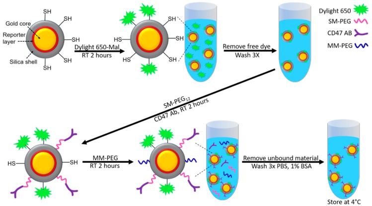
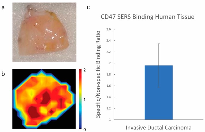
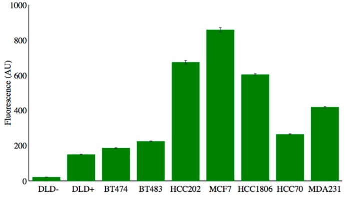
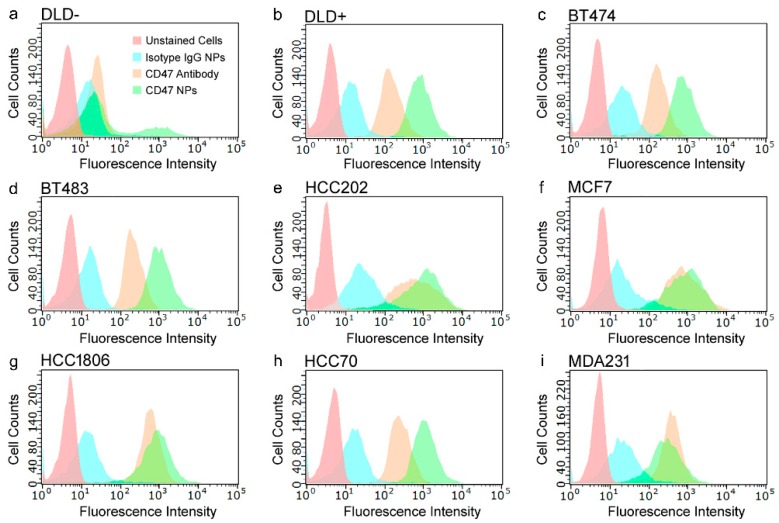
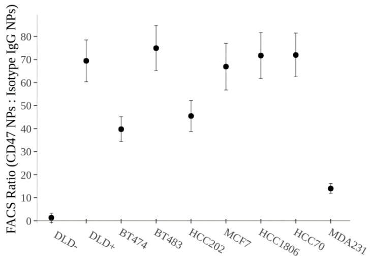
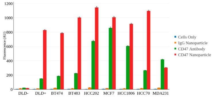
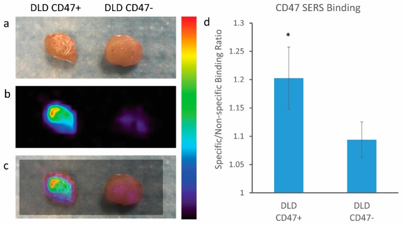
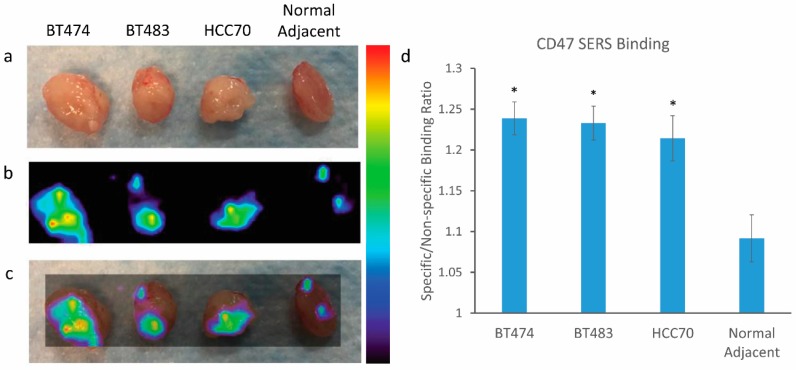
Raman spectroscopic imaging has shown great promise for improved cancer detection and localization with the use of tumor targeting surface enhanced Raman scattering (SERS) nanoparticles. With the ultrasensitive detection and multiplexing capabilities that SERS imaging has to offer, scientists have been investigating several clinical applications that could benefit from this unique imaging strategy. Recently, there has been a push to develop new image-guidance tools for surgical resection to help surgeons sensitively and specifically identify tumor margins in real time. We hypothesized that SERS nanoparticles (NPs) topically applied to breast cancer resection margins have the potential to provide real-time feedback on the presence of residual cancer in the resection margins during lumpectomy. Here, we explore the ability of SERS nanoparticles conjugated with a cluster of differentiation-47 (CD47) antibody to target breast cancer. CD47 is a cell surface receptor that has recently been shown to be overexpressed on several solid tumor types. The binding potential of our CD47-labeled SERS nanoparticles was assessed using fluorescence assisted cell sorting (FACS) on seven different human breast cancer cell lines, some of which were triple negative (negative expression of estrogen receptor (ER), progesterone receptor (PR), and human epidermal growth factor receptor-2 (HER2)). Xenograft mouse models were also used to assess the ability of our Raman imaging system to identify tumor from normal tissue. A ratiometric imaging strategy was used to quantify specific vs. nonspecific probe binding, resulting in improved tumor-to-background ratios. FACS analysis showed that CD47-labeled SERS nanoparticles bound to seven different breast cancer cell lines at levels 12-fold to 70-fold higher than isotype control-labeled nanoparticles ( < 0.01), suggesting that our CD47-targeted nanoparticles actively bind to CD47 on breast cancer cells. In a mouse xenograft model of human breast cancer, topical application of CD47-targeted nanoparticles to excised normal and cancer tissue revealed increased binding of CD47-targeted nanoparticles on tumor relative to normal adjacent tissue. The findings of this study support further investigation and suggest that SERS nanoparticles topically applied to breast cancer could guide more complete surgical resection during lumpectomy.
拉曼光谱成像在利用肿瘤靶向表面增强拉曼散射(SERS)纳米颗粒改善癌症检测和定位方面显示出巨大潜力。凭借SERS成像所具备的超灵敏检测和多重检测能力,科学家们一直在研究多种可受益于这种独特成像策略的临床应用。最近,人们一直在推动开发用于手术切除的新图像引导工具,以帮助外科医生实时灵敏且特异性地识别肿瘤边缘。我们假设,局部应用于乳腺癌切除边缘的SERS纳米颗粒(NPs)有可能在乳房肿瘤切除术期间就切除边缘是否存在残留癌提供实时反馈。在此,我们探究了与分化簇47(CD47)抗体偶联的SERS纳米颗粒靶向乳腺癌的能力。CD47是一种细胞表面受体,最近已证明在多种实体瘤类型中过表达。我们使用荧光辅助细胞分选(FACS)对七种不同的人乳腺癌细胞系评估了我们的CD47标记的SERS纳米颗粒的结合潜力,其中一些是三阴性的(雌激素受体(ER)、孕激素受体(PR)和人表皮生长因子受体2(HER2)阴性表达)。还使用异种移植小鼠模型评估了我们的拉曼成像系统从正常组织中识别肿瘤的能力。采用了一种比率成像策略来量化特异性与非特异性探针结合,从而提高了肿瘤与背景的比率。FACS分析表明,CD47标记的SERS纳米颗粒与七种不同乳腺癌细胞系的结合水平比同型对照标记的纳米颗粒高12倍至70倍(<0.01),这表明我们的CD47靶向纳米颗粒能主动结合乳腺癌细胞上的CD47。在人乳腺癌的小鼠异种移植模型中,将CD47靶向纳米颗粒局部应用于切除的正常和癌组织,结果显示相对于相邻正常组织,CD47靶向纳米颗粒在肿瘤上的结合增加。本研究结果支持进一步研究,并表明局部应用于乳腺癌的SERS纳米颗粒可在乳房肿瘤切除术期间指导更完整的手术切除。