Singapore Institute for Neurotechnology (SINAPSE), National University of Singapore, Singapore 117456, Singapore.
Department of Biomedical Engineering, National University of Singapore, Singapore 117583, Singapore.
Sensors (Basel). 2018 Nov 21;18(11):4081. doi: 10.3390/s18114081.
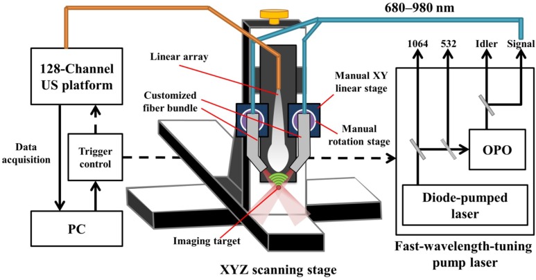
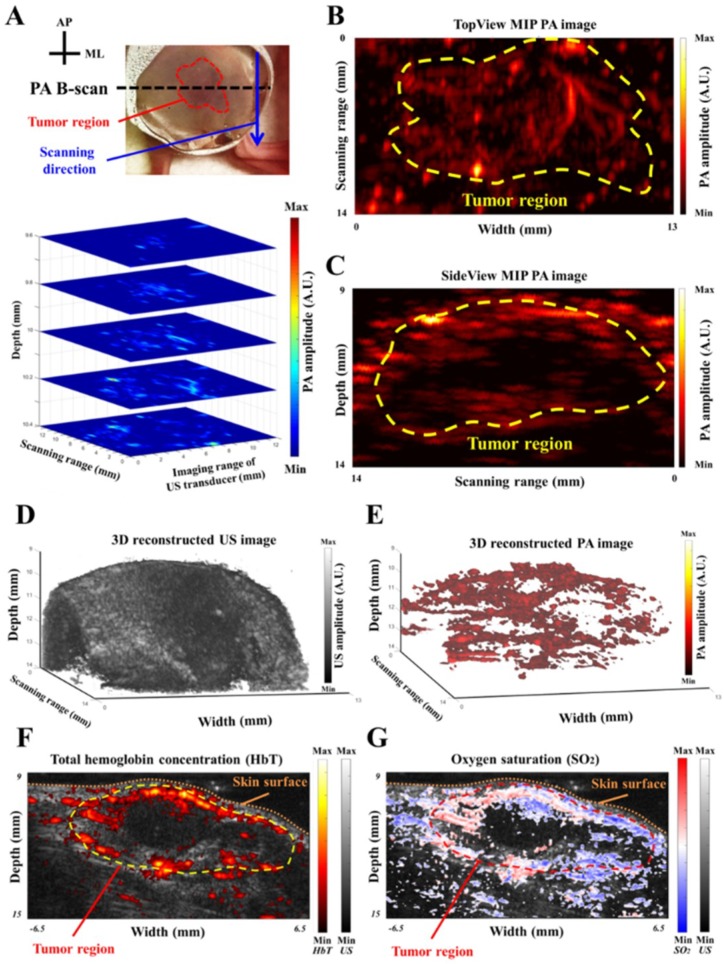
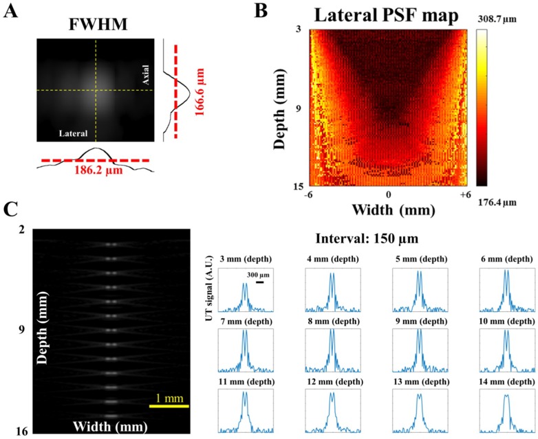
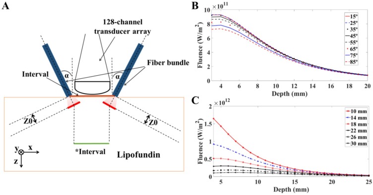
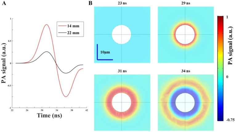
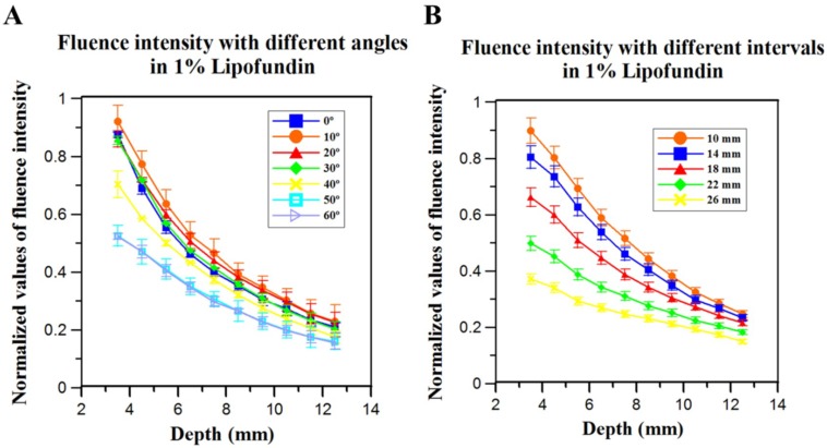
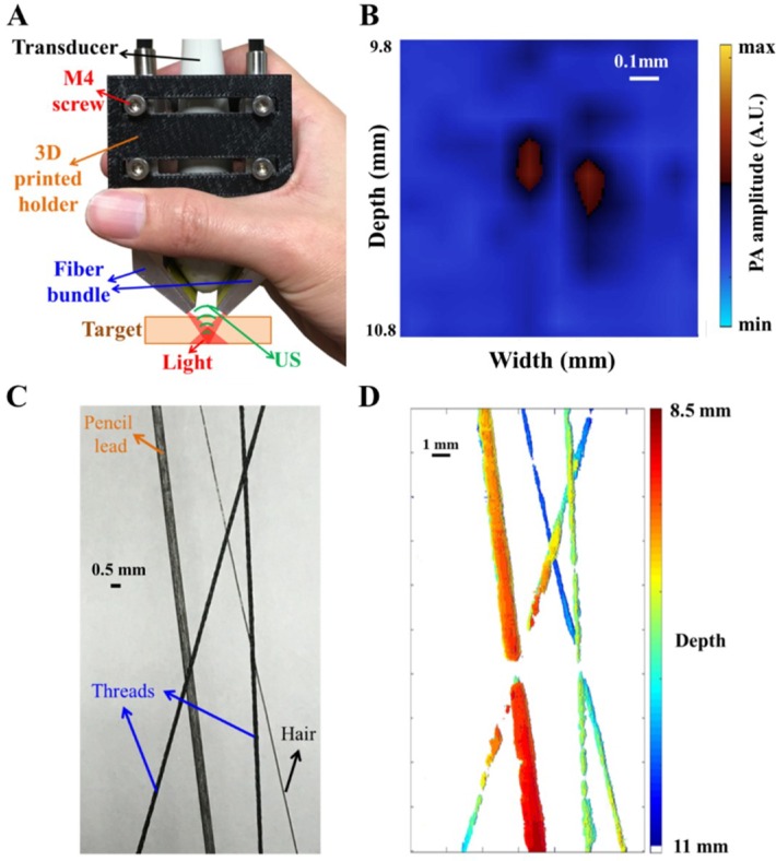
This article provides a guide to design and build a handheld, real-time photoacoustic (PA) imaging system from simulation to realization for animal neurological disease models. A pulsed laser and array-based ultrasound (US) platform were utilized to develop the system for evaluating vascular functions in rats with focal ischemia or subcutaneous tumors. To optimize the laser light delivery, finite element (FE)-based simulation models were developed to provide information regarding light propagation and PA wave generation in soft tissues. Besides, simulations were also conducted to evaluate the ideal imaging resolution of the US system. As a result, a PA C-scan image of a designed phantom in 1% Lipofundin was reconstructed with depth information. Performance of the handheld PA system was tested in an animal ischemia model, which revealed that cerebral blood volume (CBV) changes at the cortical surface could be monitored immediately after ischemia induction. Another experiment on subcutaneous tumors showed the anomalous distribution of the total hemoglobin concentration (HbT) and oxygen saturation (SO₂), while 3D and maximum intensity projection (MIP) PA images of the subcutaneous tumors are also presented in this article. Overall, this system shows promise for monitoring disease progression in vascular functional impairments.
本文提供了一个从模拟到实现的指南,用于设计和构建用于动物神经疾病模型的手持式实时光声(PA)成像系统。该系统使用脉冲激光和基于阵列的超声(US)平台来开发,用于评估局灶性缺血或皮下肿瘤大鼠的血管功能。为了优化激光光传输,开发了基于有限元(FE)的模拟模型,以提供有关软组织中光传播和 PA 波产生的信息。此外,还进行了模拟以评估 US 系统的理想成像分辨率。结果,在 1% Lipofundin 中重建了具有深度信息的设计体模的 PA C 扫描图像。在动物缺血模型中测试了手持式 PA 系统的性能,结果表明,在诱导缺血后可以立即监测皮质表面的脑血容量(CBV)变化。皮下肿瘤的另一个实验表明总血红蛋白浓度(HbT)和氧饱和度(SO₂)的异常分布,同时还展示了皮下肿瘤的 3D 和最大强度投影(MIP)PA 图像。总体而言,该系统有望用于监测血管功能障碍疾病的进展。