• 文献检索
  • 文档翻译
  • 深度研究
  • 学术资讯
  • Suppr Zotero 插件Zotero 插件
  • 邀请有礼
  • 套餐&价格
  • 历史记录
应用&插件
Suppr Zotero 插件Zotero 插件浏览器插件Mac 客户端Windows 客户端微信小程序
定价
高级版会员购买积分包购买API积分包
服务
文献检索文档翻译深度研究API 文档MCP 服务
关于我们
关于 Suppr公司介绍联系我们用户协议隐私条款
关注我们

Suppr 超能文献

核心技术专利:CN118964589B侵权必究
粤ICP备2023148730 号-1Suppr @ 2026

文献检索

告别复杂PubMed语法,用中文像聊天一样搜索,搜遍4000万医学文献。AI智能推荐,让科研检索更轻松。

立即免费搜索

文件翻译

保留排版,准确专业,支持PDF/Word/PPT等文件格式,支持 12+语言互译。

免费翻译文档

深度研究

AI帮你快速写综述,25分钟生成高质量综述,智能提取关键信息,辅助科研写作。

立即免费体验

多发性硬化症中多灶性视觉诱发电位和对比敏感度与节细胞-内丛状层厚度相关。

Multifocal visual evoked potentials and contrast sensitivity correlate with ganglion cell-inner plexiform layer thickness in multiple sclerosis.

机构信息

College of Optometry, University of Houston, Houston, TX, USA.

College of Optometry, University of Houston, Houston, TX, USA.

出版信息

Clin Neurophysiol. 2019 Jan;130(1):180-188. doi: 10.1016/j.clinph.2018.10.007. Epub 2018 Nov 13.

DOI:10.1016/j.clinph.2018.10.007
PMID:30473445
原文链接:https://pmc.ncbi.nlm.nih.gov/articles/PMC6309727/
Abstract

OBJECTIVE

To examine the relationship between optical coherence tomography (OCT) macular ganglion cell-inner plexiform layer thickness (GCIPLT), peripapillary retinal nerve fiber layer thickness (RNFLT) and visual function in relapsing-remitting multiple sclerosis (RRMS).

METHODS

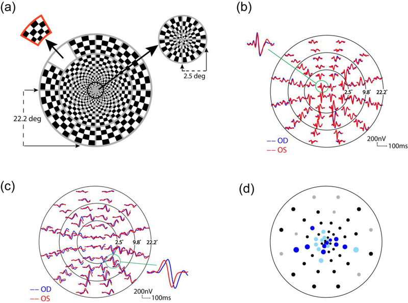
Cirrus OCT, VERIS 60-sector multifocal visual evoked potential (mfVEP) and Pelli-Robson contrast sensitivity (CS) were obtained for 53 eyes with last optic neuritis (ON) > 6 months and 105 non-ON eyes in 90 patients. One eye (43 ON, 73 non-ON) was used for correlations when both had the same history. Global (G, 60 sectors) and central 5.6° (C, 24 sectors) mfVEP amplitude and latency were calculated as mean logSNR and median latency.

RESULTS

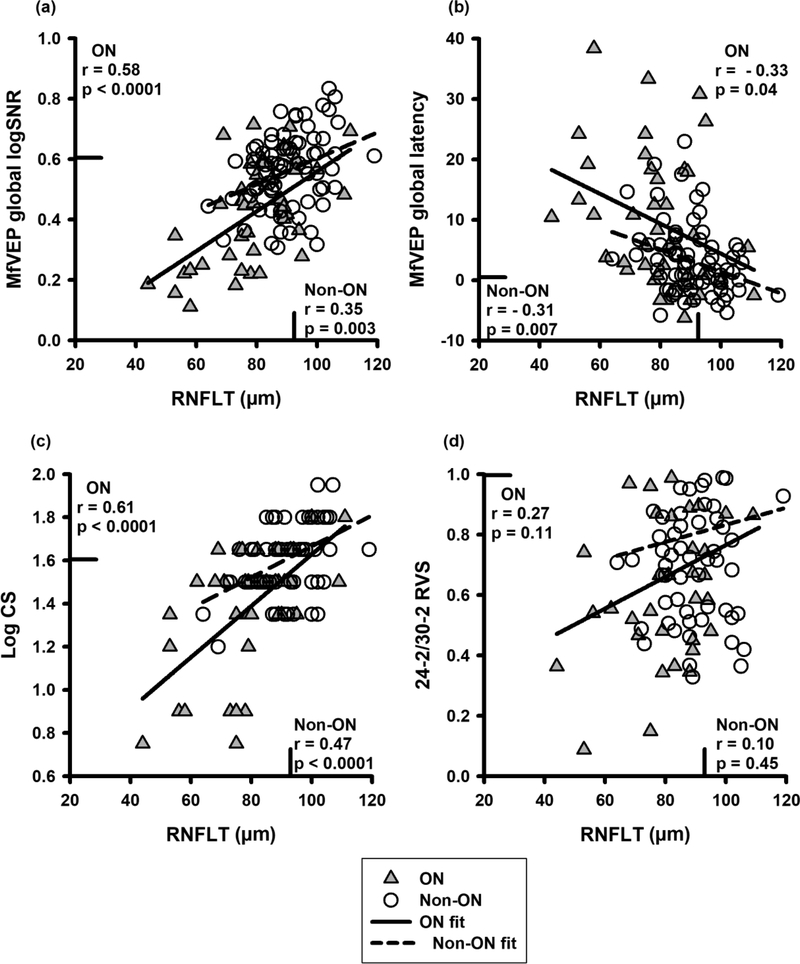
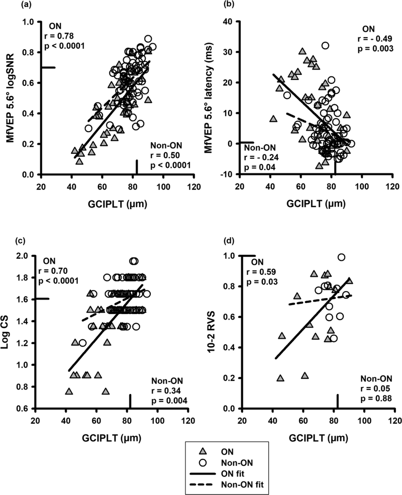
Eyes showing abnormal mfVEP (amplitude or latency) vs OCT (GCIPLT or RNFLT) was 77% vs 69% (p = 0.33) in ON, 45% vs 22% (p < 0.0005) in non-ON. In ON and non-ON, mfVEP measures and CS correlated with GCIPLT and RNFLT (r = -0.24 to 0.78, p = 0.03-0.0001). In ON, mfVEP amplitude (C,G) correlated better with GCIPLT (r = 0.78, 0.76) than RNFLT (r = 0.43, 0.58; p < 0.001, 0.01).

CONCLUSIONS

MfVEP measures and CS correlated well with GCIPLT and RNFLT in ON and non-ON. MfVEP amplitudes were more highly correlated with GCIPLT than RNFLT in ON. MfVEP detected significantly more defects than OCT in non-ON.

SIGNIFICANCE

GCIPLT, mfVEP and CS provide useful measures of optic nerve integrity in RRMS.

摘要

目的

探讨光学相干断层扫描(OCT)黄斑神经节细胞-内丛状层厚度(GCIPLT)、视盘周围视网膜神经纤维层厚度(RNFLT)与复发缓解型多发性硬化症(RRMS)患者视力的关系。

方法

对 90 例患者的 53 只曾患视神经炎(ON)眼(ON 眼)和 105 只未患视神经炎眼(非 ON 眼)进行 Cirrus OCT、VERIS 60 区多焦视觉诱发电位(mfVEP)和 Pelli-Robson 对比敏感度(CS)检查。当两只眼具有相同病史时,使用其中一只眼(43 只 ON 眼和 73 只非 ON 眼)进行相关性分析。计算平均对数信噪比(logSNR)和中位数潜伏期作为整体(G,60 区)和中央 5.6°(C,24 区)mfVEP 的振幅和潜伏期。

结果

ON 眼和非 ON 眼中,mfVEP 检查(振幅或潜伏期)异常而 OCT 检查(GCIPLT 或 RNFLT)正常的比例分别为 77%和 69%(p=0.33);mfVEP 检查异常而 OCT 检查正常的比例分别为 45%和 22%(p<0.0005)。ON 眼和非 ON 眼中,mfVEP 测量值和 CS 与 GCIPLT 和 RNFLT 相关(r=-0.24 至 0.78,p=0.03-0.0001)。在 ON 眼,mfVEP 振幅(C,G)与 GCIPLT 的相关性优于 RNFLT(r=0.78,0.76 比 r=0.43,0.58;p<0.001,0.01)。

结论

ON 眼和非 ON 眼中,mfVEP 测量值和 CS 与 GCIPLT 和 RNFLT 相关性良好。在 ON 眼,mfVEP 振幅与 GCIPLT 的相关性优于 RNFLT。非 ON 眼中,mfVEP 比 OCT 检测到更多的异常。

意义

GCIPLT、mfVEP 和 CS 为 RRMS 视神经完整性提供了有用的测量指标。

https://cdn.ncbi.nlm.nih.gov/pmc/blobs/03e9/6309727/7c31fe8189c3/nihms-1514307-f0003.jpg
https://cdn.ncbi.nlm.nih.gov/pmc/blobs/03e9/6309727/f5b0870d8a00/nihms-1514307-f0001.jpg
https://cdn.ncbi.nlm.nih.gov/pmc/blobs/03e9/6309727/7b2f1d3ac757/nihms-1514307-f0002.jpg
https://cdn.ncbi.nlm.nih.gov/pmc/blobs/03e9/6309727/7c31fe8189c3/nihms-1514307-f0003.jpg
https://cdn.ncbi.nlm.nih.gov/pmc/blobs/03e9/6309727/f5b0870d8a00/nihms-1514307-f0001.jpg
https://cdn.ncbi.nlm.nih.gov/pmc/blobs/03e9/6309727/7b2f1d3ac757/nihms-1514307-f0002.jpg
https://cdn.ncbi.nlm.nih.gov/pmc/blobs/03e9/6309727/7c31fe8189c3/nihms-1514307-f0003.jpg

相似文献

1
Multifocal visual evoked potentials and contrast sensitivity correlate with ganglion cell-inner plexiform layer thickness in multiple sclerosis.多发性硬化症中多灶性视觉诱发电位和对比敏感度与节细胞-内丛状层厚度相关。
Clin Neurophysiol. 2019 Jan;130(1):180-188. doi: 10.1016/j.clinph.2018.10.007. Epub 2018 Nov 13.
2
Differing Structural and Functional Patterns of Optic Nerve Damage in Multiple Sclerosis and Neuromyelitis Optica Spectrum Disorder.多发性硬化症和视神经脊髓炎谱系障碍视神经损伤的结构和功能模式差异。
Ophthalmology. 2019 Mar;126(3):445-453. doi: 10.1016/j.ophtha.2018.06.022. Epub 2018 Jul 27.
3
Evaluation of visual structural and functional factors that predict the development of multiple sclerosis in clinically isolated syndrome patients.评估预测临床孤立综合征患者多发性硬化症发生发展的视觉结构和功能因素。
Invest Ophthalmol Vis Sci. 2014 Sep 4;55(10):6127-31. doi: 10.1167/iovs.14-14807.
4
Functional-structural assessment of the optic pathways in patients with optic neuritis.视神经炎患者视路的功能-结构评估
Doc Ophthalmol. 2020 Apr;140(2):159-168. doi: 10.1007/s10633-019-09728-0. Epub 2019 Oct 17.
5
Comparison of multifocal visual evoked potential, standard automated perimetry and optical coherence tomography in assessing visual pathway in multiple sclerosis patients.比较多焦视觉诱发电位、标准自动视野计和光学相干断层扫描在评估多发性硬化症患者视路中的应用。
Mult Scler. 2010 Apr;16(4):412-26. doi: 10.1177/1352458509359782. Epub 2010 Mar 5.
6
Longitudinal Evaluation of Visual Function in Multiple Sclerosis.多发性硬化症视觉功能的纵向评估
Optom Vis Sci. 2015 Oct;92(10):976-85. doi: 10.1097/OPX.0000000000000684.
7
Reproducibility of multifocal visual evoked potential and traditional visual evoked potential in normal and multiple sclerosis eyes.正常及多发性硬化症患者眼中多焦视觉诱发电位和传统视觉诱发电位的可重复性
Doc Ophthalmol. 2015 Feb;130(1):31-41. doi: 10.1007/s10633-014-9467-5. Epub 2014 Oct 29.
8
Multifocal Visual Evoked Potential in Eyes With Temporal Hemianopia From Chiasmal Compression: Correlation With Standard Automated Perimetry and OCT Findings.视交叉受压所致颞侧偏盲眼中的多焦视觉诱发电位:与标准自动视野计及光学相干断层扫描结果的相关性
Invest Ophthalmol Vis Sci. 2017 Sep 1;58(11):4436-4449. doi: 10.1167/iovs.17-21529.
9
Evaluation of the Innermost Retinal Layers and Visual Evoked Potentials in Patients with Multiple Sclerosis.多发性硬化症患者最内层视网膜层及视觉诱发电位的评估
Curr Eye Res. 2016 Oct;41(10):1353-1358. doi: 10.3109/02713683.2015.1119283. Epub 2016 Feb 16.
10
Relationship between optical coherence tomography and electrophysiology of the visual pathway in non-optic neuritis eyes of multiple sclerosis patients.多发性硬化症患者非视神经炎眼中光学相干断层扫描与视觉通路电生理学之间的关系
PLoS One. 2014 Aug 28;9(8):e102546. doi: 10.1371/journal.pone.0102546. eCollection 2014.

引用本文的文献

1
Evaluation of Retinal Inner Layer Thickness and its Relationship with Visual Prognosis in Multiple Sclerosis Patients with and Without Optic Neuritis.多发性硬化症伴或不伴视神经炎患者视网膜内层厚度评估及其与视觉预后的关系
Beyoglu Eye J. 2025 Jun 25;10(2):95-100. doi: 10.14744/bej.2025.48379. eCollection 2025.
2
Association Between Contrast Sensitivity and Ganglion Cell-Inner Plexiform Layer Thickness After Resolution of Macular Edema Due to Branch Retinal Vein Occlusion.视网膜分支静脉阻塞所致黄斑水肿消退后对比敏感度与神经节细胞-内丛状层厚度之间的关联
J Clin Med. 2025 Feb 24;14(5):1507. doi: 10.3390/jcm14051507.
3

本文引用的文献

1
Translating data and measurements from stratus to cirrus OCT in glaucoma patients and healthy subjects.在青光眼患者和健康受试者中,将Stratus OCT的数据和测量结果转换为Cirrus OCT的数据和测量结果。
Rom J Ophthalmol. 2016 Jul-Sep;60(3):158-164.
2
No evidence of disease activity is associated with reduced rate of axonal retinal atrophy in MS.在多发性硬化症中,无疾病活动迹象与视网膜轴突萎缩速率降低相关。
Neurology. 2017 Dec 12;89(24):2469-2475. doi: 10.1212/WNL.0000000000004736. Epub 2017 Nov 15.
3
OCT as a window to the MS brain: The view becomes slightly clearer.
OCT-Based Retina Assessment Reflects Visual Impairment in Multiple Sclerosis.
基于光学相干断层扫描的视网膜评估反映了多发性硬化症中的视力损害。
Invest Ophthalmol Vis Sci. 2025 Feb 3;66(2):39. doi: 10.1167/iovs.66.2.39.
4
The Evolvement of OCT and OCT-A in Identifying Multiple Sclerosis Biomarkers.光学相干断层扫描(OCT)及光学相干断层扫描血管造影(OCT-A)在识别多发性硬化生物标志物方面的进展
Biomedicines. 2023 Nov 11;11(11):3031. doi: 10.3390/biomedicines11113031.
5
Regional Analysis of Inner Retinal Layer Changes in Multiple Sclerosis with and without Optic Neuritis.多发性硬化症伴或不伴视神经炎的内视网膜层变化的区域分析。
Turk J Ophthalmol. 2023 Jun 21;53(3):169-174. doi: 10.4274/tjo.galenos.2023.81486.
6
Macular Ganglion Cell-Inner Plexiform Layer as a Marker of Cognitive and Sensory Function in Midlife.黄斑神经节细胞-内丛状层作为中年认知和感觉功能的标志物。
J Gerontol A Biol Sci Med Sci. 2020 Sep 16;75(9):e42-e48. doi: 10.1093/gerona/glaa135.
光学相干断层扫描作为了解多发性硬化症脑部的窗口:视野变得稍清晰一些。
Neurology. 2017 Dec 12;89(24):2404-2405. doi: 10.1212/WNL.0000000000004755. Epub 2017 Nov 15.
4
The loss of macular ganglion cells begins from the early stages of disease and correlates with brain atrophy in multiple sclerosis patients.黄斑神经节细胞的丢失始于疾病的早期阶段,并与多发性硬化症患者的脑萎缩相关。
Mult Scler. 2019 Jan;25(1):31-38. doi: 10.1177/1352458517740214. Epub 2017 Nov 10.
5
Retinal layer segmentation in multiple sclerosis: a systematic review and meta-analysis.多发性硬化症中的视网膜层分割:系统评价和荟萃分析。
Lancet Neurol. 2017 Oct;16(10):797-812. doi: 10.1016/S1474-4422(17)30278-8. Epub 2017 Sep 12.
6
Retinal ganglion cell analysis in multiple sclerosis and optic neuritis: a systematic review and meta-analysis.多发性硬化症和视神经炎中的视网膜神经节细胞分析:系统评价和荟萃分析。
J Neurol. 2017 Sep;264(9):1837-1853. doi: 10.1007/s00415-017-8531-y. Epub 2017 May 31.
7
Multifocal visual evoked potentials in optic neuritis and multiple sclerosis: A review.视神经炎和多发性硬化中的多焦视觉诱发电位:综述
Clin Neurophysiol. 2017 Jul;128(7):1234-1245. doi: 10.1016/j.clinph.2017.03.047. Epub 2017 Apr 11.
8
Validity of low-contrast letter acuity as a visual performance outcome measure for multiple sclerosis.低对比度字母视力作为多发性硬化症视觉功能结果测量指标的有效性。
Mult Scler. 2017 Apr;23(5):734-747. doi: 10.1177/1352458517690822. Epub 2017 Feb 16.
9
Multiple sclerosis seen through new eyes.以新视角看待多发性硬化症。
Clin Exp Ophthalmol. 2017 Jan;45(1):9-11. doi: 10.1111/ceo.12885.
10
Quantitative visual tests after poorly recovered optic neuritis due to multiple sclerosis.多发性硬化导致的视神经炎恢复不佳后的定量视觉测试。
Mult Scler Relat Disord. 2016 Nov;10:198-203. doi: 10.1016/j.msard.2016.10.009. Epub 2016 Nov 4.