University of Houston, College of Optometry, Houston, Texas 77204-2020, USA.
Mult Scler. 2010 Apr;16(4):412-26. doi: 10.1177/1352458509359782. Epub 2010 Mar 5.
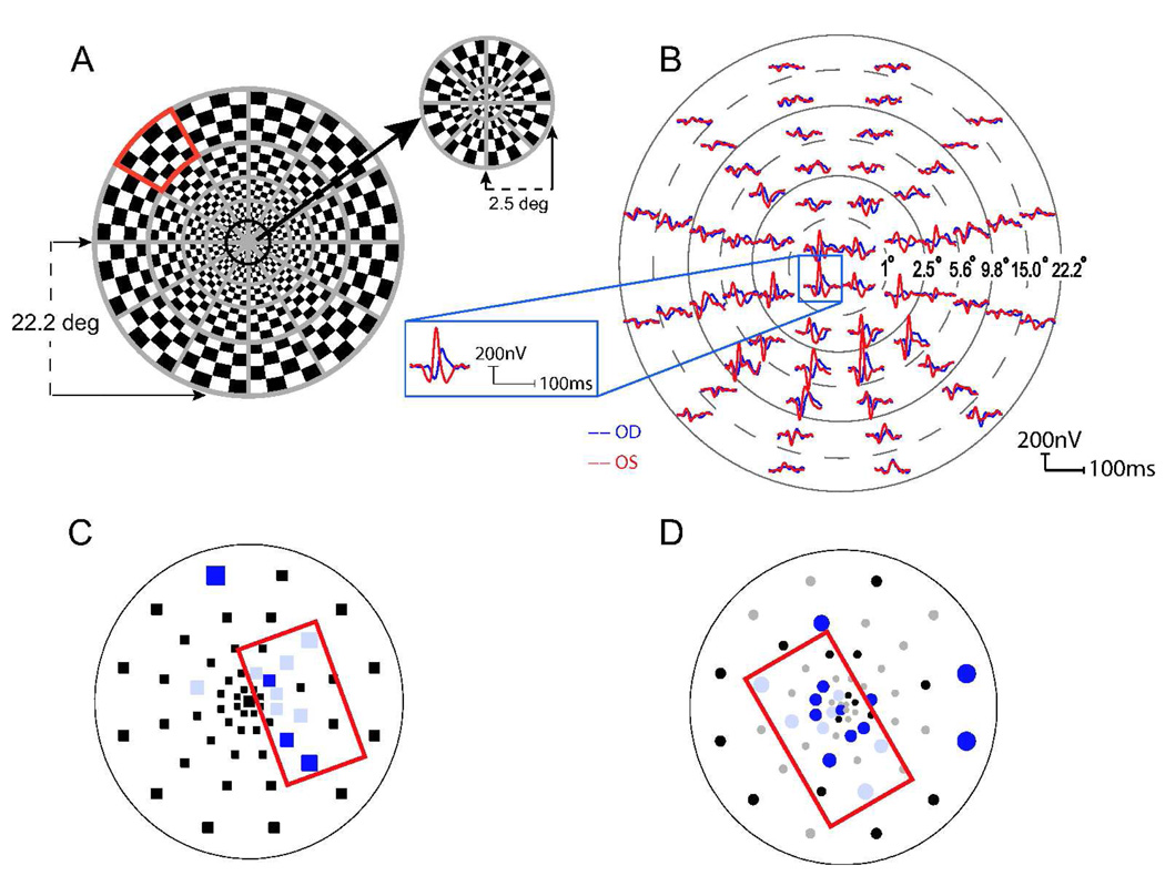
Multifocal visual evoked potentials (mfVEP) measure local response amplitude and latency in the field of vision.
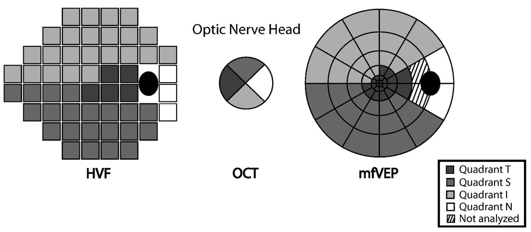
To compare the sensitivity of mfVEP, Humphrey visual field (HVF) and optical coherence tomography (OCT) in detecting visual abnormality in multiple sclerosis (MS) patients.
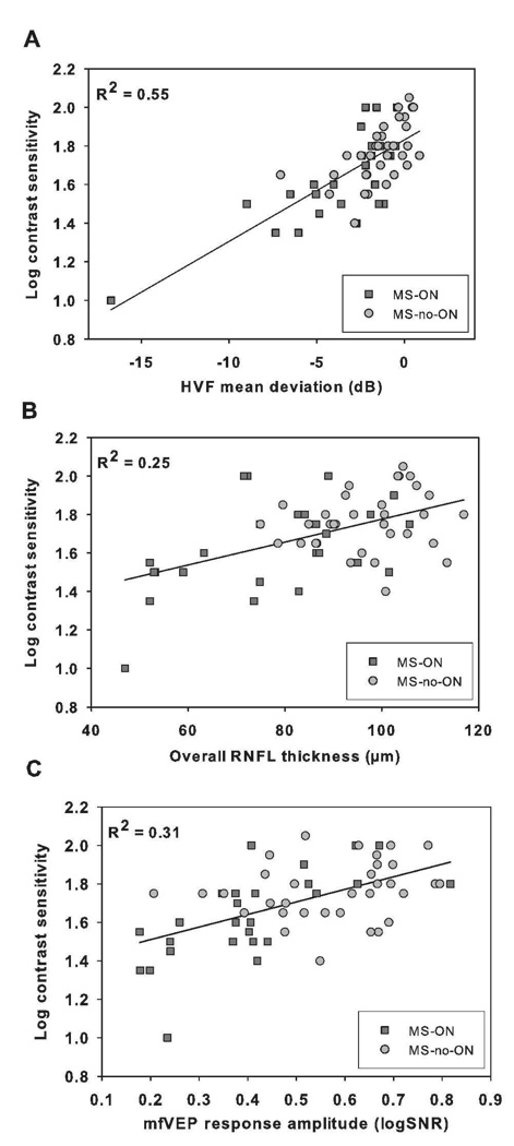
mfVEP, HVF, and OCT (retinal nerve fiber layer [RNFL]) were performed in 47 MS-ON eyes (last optic neuritis [ON] attack >or=6 months prior) and 65 MS-no-ON eyes without ON history. Criteria to define an eye as abnormal were: (1) mfVEP amplitude/latency - either amplitude or latency probability plots meeting cluster criteria with 95% specificity; (2) mfVEP amplitude or latency alone (specificity: 97% and 98%, respectively); and (3) HVF and OCT, mean deviation and RNFL thickness meeting p < 0.05, respectively.
MfVEP (amplitude/latency) identified more abnormality in MS-ON eyes (89%) than HVF (72%), OCT (62%), mfVEP amplitude (66%) or latency (67%) alone. Eighteen percent of MS-no-ON eyes were abnormal for both mfVEP (amplitude/latency) and HVF compared with 8% with OCT. Agreement between tests ranged from 60% to 79%. mfVEP (amplitude/latency) categorized an additional 15% of MS-ON eyes as abnormal compared with HVF and OCT combined.
mfVEP, which detects both demyelination (increased latency) and neural degeneration (reduced amplitude), revealed more abnormality than HVF or OCT in MS patients.
多焦视觉诱发电位(mfVEP)测量视野中的局部反应幅度和潜伏期。
比较 mfVEP、Humphrey 视野(HVF)和光学相干断层扫描(OCT)在检测多发性硬化症(MS)患者视觉异常中的敏感性。
对 47 例 MS-ON 眼(最近的视神经炎[ON]发作> = 6 个月前)和 65 例无 ON 病史的 MS-no-ON 眼进行 mfVEP、HVF 和 OCT(视网膜神经纤维层[RNFL])检查。定义异常眼的标准为:(1)mfVEP 幅度/潜伏期-要么幅度或潜伏期概率图满足 95%特异性的聚类标准;(2)mfVEP 幅度或潜伏期单独(特异性分别为 97%和 98%);(3)HVF 和 OCT,平均偏差和 RNFL 厚度分别满足 p < 0.05。
mfVEP(幅度/潜伏期)在 MS-ON 眼中(89%)比 HVF(72%)、OCT(62%)、mfVEP 幅度(66%)或潜伏期(67%)单独更能识别异常。18%的 MS-no-ON 眼 mfVEP(幅度/潜伏期)和 HVF 均异常,而 OCT 为 8%。测试之间的一致性范围为 60%至 79%。与 HVF 和 OCT 联合使用相比,mfVEP(幅度/潜伏期)还将另外 15%的 MS-ON 眼归类为异常。
mfVEP 可检测脱髓鞘(潜伏期增加)和神经退行性变(幅度降低),在 MS 患者中比 HVF 或 OCT 更能发现异常。