Rana Mrinal, Shah Sunil, Pandey Pravin, Masood Imran
Consultant, Department of Ophthalmology, Peterborough City Hospital, North West Anglia Hospital NHS Trust, Bretton Gate, Peterborough, UK.
Lead Consultant, Department of Ophthalmology, Birmingham and Midland Eye Centre, UK.
J Curr Glaucoma Pract. 2018 May-Aug;12(2):90-93. doi: 10.5005/jp-journals-10008-1250. Epub 2018 Aug 1.
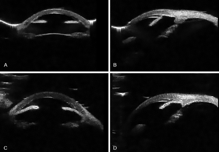
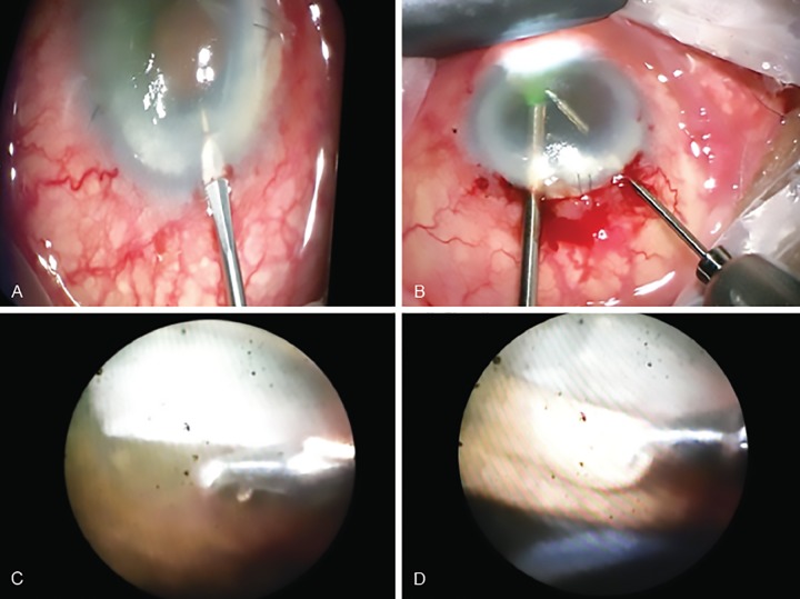
We describe a new modified technique to release the peripheral iridocorneal adhesions that formed after Descemet stripping automated endothelial keratoplasty. The usual technique of goniosynechialysis was modified and performed using endoscopic fiber-optic light and camera probe to aid visualization of the adherent iris tissue and carry out uneventful 270 degrees release of adhesions. The iris tissue was gently pulled away using micro forceps. The modified technique was conceptualized, as the view from the cornea was very poor due to recent lamellar surgery and corneal oedema secondary to poorly controlled intraocular pressure. The blocked trabecular meshwork system was successfully recanalized, which allowed an adequate control of intraocular pressure. The graft survived the insult and cornea gained complete clarity giving the patient the desired vision and improved quality of life. Rana M, Shah S, Pandey P, Masood I. Endoscopic Goniosynechialysis for Acute Angle Closure Glaucoma Following Descemet's Stripping Automated Endothelial Keratoplasty. J Curr Glaucoma Pract 2018;12(2):90-93.
我们描述了一种新的改良技术,用于松解在Descemet膜剥脱自动内皮角膜移植术后形成的周边虹膜角膜粘连。改良了通常的房角粘连分离术,使用内镜光纤照明和摄像探头辅助观察粘连的虹膜组织,并顺利进行270度的粘连松解。使用微型镊子轻轻拉开虹膜组织。由于近期的板层手术以及眼压控制不佳继发角膜水肿,角膜视野很差,因此构思了这种改良技术。成功使阻塞的小梁网系统再通,从而实现了对眼压的充分控制。移植片经受住了损伤,角膜完全恢复透明,使患者获得了理想的视力并改善了生活质量。Rana M、Shah S、Pandey P、Masood I。Descemet膜剥脱自动内皮角膜移植术后急性闭角型青光眼的内镜下房角粘连分离术。《当代青光眼实践杂志》2018年;12(2):90-93。