Zhang Junyu, Lu Yan, Hsi Wenchien, Zhang Jiangang, Sheng Yinxiangzi, Shi Leijun, Wang Weiwei, Lu Jiade, Zhou Rong, Cheng Jingyi
College of Physical Science and Technology (College of Nuclear Science and Engineering), Sichuan University, Chengdu, China.
Key Laboratory of Radiation Physics and Technology Ministry of Education, Chengdu, China.
Front Oncol. 2018 Nov 13;8:523. doi: 10.3389/fonc.2018.00523. eCollection 2018.
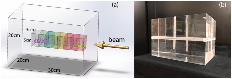
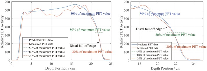
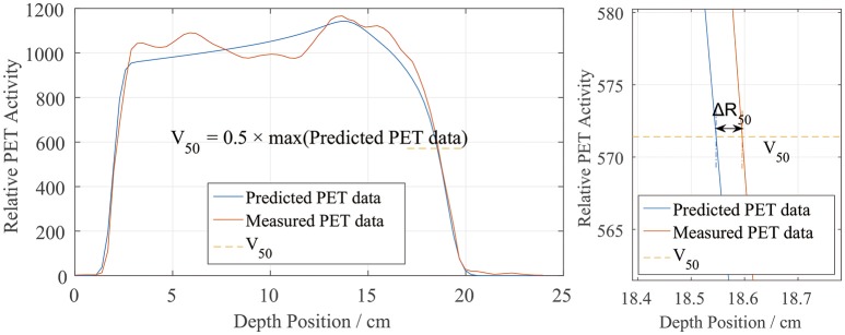
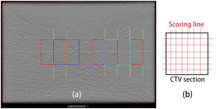
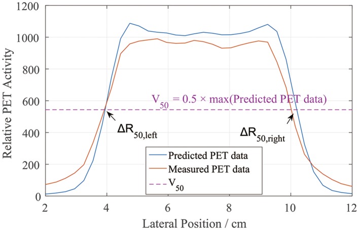
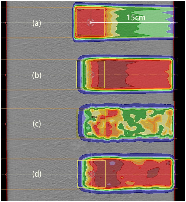
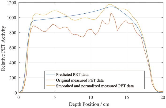
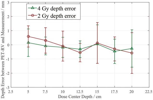
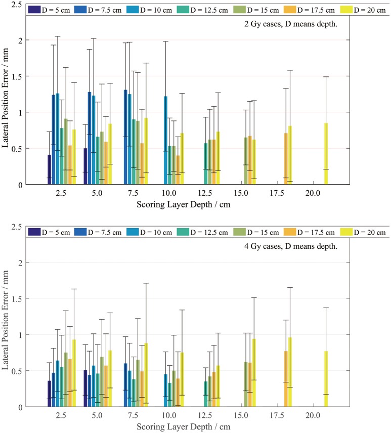
Positron emission tomography (PET) scanning is a widely used method of proton therapy verification. In this study, a proton radiotherapy accuracy verification process was developed by comparing predicted and measured PET data to verify the correctness of PET prediction and was tested at the Shanghai Proton and Heavy Ion Center. Irradiation was performed on a polymethyl methacrylate (PMMA) phantom. There were two dose groups, to which 2 and 4 Gy doses were delivered, and each dose group had different designed dose depths ranging from 5 to 20 cm. The predicted PET results were obtained using a PET prediction calculation module. The measured data were collected with a PET/computed tomography device. The predicted and measured PET data were normalized to similar PET amplitude values before comparison and were compared using depth and lateral profiles for the position error. The error was evaluated at the position corresponding to 50% of the maximum on the PET curves. The mean and standard deviation were calculated based on the data sampled in the scoring area. Gamma index analysis is also applied in the comparison. In the depth comparison, the 2 and 4 Gy dose cases yielded similar mean depth errors between 1 and -1 mm, and the deviation was <2 mm. In the lateral comparison, the 2 Gy cases had a mean lateral error around 1 mm, and the 4 Gy cases had a mean lateral error <1 mm, with a standard deviation <1 mm for both the 2 and 4 Gy cases. All the cases have a gamma passing rate over 95%. The comparison of these PMMA phantom cases revealed good agreement between the predicted and measured PET data, with depth and lateral position errors <2 mm in total, considering the uncertainty. The comparison results demonstrate that the PET predictions obtained in PMMA phantom tests for single proton beam therapy verification are reliable and that the research can be extended to verification in human body treatment with further investigation.
正电子发射断层扫描(PET)是一种广泛应用于质子治疗验证的方法。在本研究中,通过比较预测的和测量的PET数据,开发了一种质子放射治疗准确性验证流程,以验证PET预测的正确性,并在上海质子重离子中心进行了测试。在聚甲基丙烯酸甲酯(PMMA)体模上进行照射。有两个剂量组,分别给予2 Gy和4 Gy的剂量,每个剂量组有不同的设计剂量深度,范围为5至20 cm。使用PET预测计算模块获得预测的PET结果。用PET/计算机断层扫描设备收集测量数据。在比较之前,将预测的和测量的PET数据归一化为相似的PET幅度值,并使用深度和横向剖面比较位置误差。在PET曲线上最大值的50%对应的位置评估误差。根据在评分区域采样的数据计算平均值和标准差。比较中还应用了伽马指数分析。在深度比较中,2 Gy和4 Gy剂量的情况产生的平均深度误差在1至 -1 mm之间,偏差<2 mm。在横向比较中,2 Gy剂量的情况平均横向误差约为1 mm,4 Gy剂量的情况平均横向误差<1 mm,2 Gy和4 Gy剂量的情况标准差均<1 mm。所有情况的伽马通过率均超过95%。这些PMMA体模情况的比较表明,预测的和测量的PET数据之间具有良好的一致性,考虑到不确定性,深度和横向位置误差总计<2 mm。比较结果表明,在PMMA体模测试中获得的用于单质子束治疗验证的PET预测是可靠的,并且该研究可以通过进一步研究扩展到人体治疗的验证。